
Dr. Samhitha Reddy
Consultant Proctologist & Laparoscopic Surgeon
MBBS, MS, FMAS, FISCP, DMAS
- 8 Yrs
- ASCRS Member
Persistent upper neck pain, difficulty turning the head, or worsening balance and coordination problems may be associated with Atlanto-Axial Instability (AAI). This condition occurs when the joint between the first and second cervical vertebrae becomes abnormally unstable. The C1–C2 region supports the head and allows the neck to rotate, and instability here can occasionally compress the spinal cord and cause serious neurological complications.
Treatment depends on the cause, the degree of instability and whether the spinal cord is at risk. Some carefully selected patients may undergo observation, while others require surgical stabilisation. At Lux Hospitals, Hyderabad, patients can enquire about specialist spine consultations for suspected upper cervical instability and related neurological symptoms.
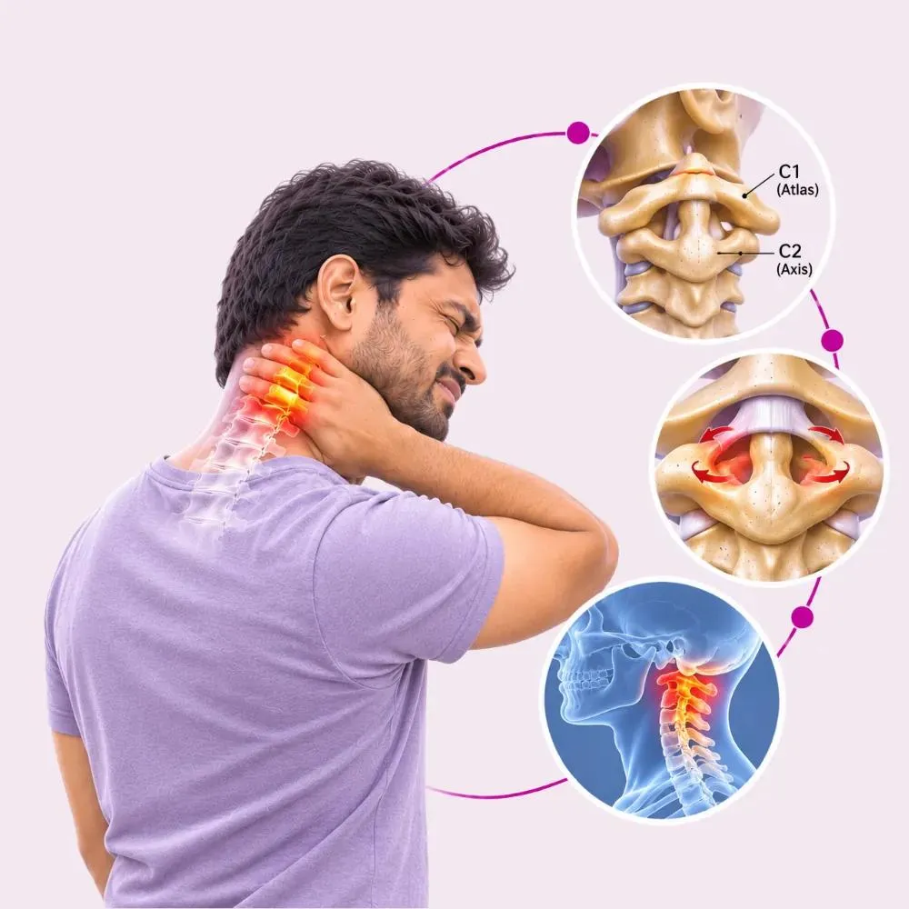
Atlanto-axial instability is a condition involving abnormal movement or reduced stability between the first cervical vertebra (atlas, C1) and the second cervical vertebra (axis, C2). These vertebrae form the atlanto-axial joint, which allows head rotation.
Strong ligaments, particularly the transverse ligament, help maintain their alignment and stability. When these supporting structures are damaged, weakened or abnormally developed, excessive movement may occur, potentially narrowing the space available for the spinal cord.
Atlanto-axial instability may be associated with abnormal vertebral alignment, known as atlanto-axial subluxation or dislocation. Significant instability can compress the spinal cord, causing neurological symptoms and, in severe cases, serious complications.
Although some people may have radiological evidence of instability without noticeable symptoms, confirmed cases require specialist evaluation to assess spinal stability and determine appropriate treatment.
Atlanto-axial instability may result from congenital abnormalities, trauma, inflammatory disease or damage to the supporting ligaments.
Some people are born with structural or developmental differences in the C1–C2 vertebrae. These abnormalities may increase susceptibility to instability.
Os odontoideum is a condition in which the odontoid process is separated from the body of the axis. It may be associated with C1–C2 instability and, in selected cases, spinal cord compression.
Rheumatoid arthritis can affect the joints and ligaments of the upper cervical spine. Chronic inflammation may damage stabilising structures and contribute to atlanto-axial subluxation.
Road accidents, falls and other injuries can damage the ligaments or bones supporting the C1–C2 joint. Traumatic instability requires urgent assessment and appropriate cervical spine protection.
Damage to the transverse ligament or other supporting structures can reduce joint stability.
Some people with Down syndrome or selected skeletal dysplasias may have an increased risk of atlanto-axial instability. However, these conditions do not automatically mean that clinically significant instability is present.
Rarely, inflammatory conditions affecting the upper cervical region may produce abnormal alignment or instability. Grisel syndrome is one example of non-traumatic atlanto-axial subluxation linked to inflammation.
The cause matters for treatment: instability from a congenital defect or rheumatoid arthritis may not be managed in the same way as traumatic instability.
Symptoms range from mild upper neck discomfort to significant neurological impairment. Some patients have radiological instability without noticeable symptoms.
Common symptoms
Emergency warning signs
These symptoms are not specific to atlanto-axial instability and may occur with several other cervical conditions. Do not attempt neck manipulation, forceful stretching or self-directed repositioning when upper cervical instability is suspected. After significant trauma, avoid unnecessary neck movement until a qualified trauma team has assessed the cervical spine.
Diagnosis requires assessment of upper cervical anatomy, neurological function and the stability of the C1–C2 joint.
The specialist reviews neck symptoms, previous injuries, rheumatoid arthritis, congenital conditions and neurological complaints. The timing and progression of symptoms are particularly important.
The specialist may assess muscle strength, sensation, reflexes, walking ability and coordination. The examination helps identify possible spinal cord dysfunction.
Appropriate cervical radiographs may show C1–C2 alignment. An open-mouth odontoid view or a lateral cervical view may be used, depending on the clinical situation.
CT provides detailed images of the bones of the upper cervical spine. It may help identify fractures, congenital abnormalities, os odontoideum and abnormal vertebral alignment, and CT with three-dimensional reconstruction may also support surgical planning.
MRI helps assess the spinal cord, ligaments and surrounding soft tissues. It is particularly important when neurological deficits or spinal cord compression are suspected.
The specialist reviews the relationship between C1 and C2 and determines whether instability is present. Measurements such as the atlantodental interval and the space available for the spinal cord may be considered.
The findings are reviewed together to decide whether instability is present, whether the spinal cord is at risk and which treatment is appropriate.
Treatment depends on the cause of instability, spinal alignment, symptoms and the presence of neurological impairment. Selected patients without significant neurological compromise or high-risk instability may be managed through specialist observation.
Regular specialist assessments may help identify changes in neurological symptoms or function.
Selected patients may require repeat imaging to assess alignment or progression. Imaging type and timing are individualised.
An appropriately selected cervical orthosis may be recommended in certain injuries or specific clinical situations. The type of immobilisation and treatment duration depend on the diagnosis. A soft collar is not an adequate substitute for treatment of significant mechanical instability.
Patients with rheumatoid arthritis or other associated diseases may need treatment for the underlying condition alongside spine assessment.
The treating specialist may recommend avoiding contact sports, high-impact activity or movements that place excessive stress on the upper cervical spine.
Rehabilitation may be appropriate for selected patients only after stability has been established and the treating specialist has confirmed which movements are safe. Physiotherapy does not mechanically correct significant C1–C2 instability, and forceful cervical manipulation, traction or aggressive neck exercises should not be used without specialist assessment and explicit clearance.
Stabilises the atlas and axis using appropriate implants and bone grafting. It is a common surgical approach for selected cases of atlanto-axial instability.
Specialised screws and connecting rods may stabilise the C1–C2 joint. Implant selection depends on anatomy, bone quality and the location of nearby blood vessels.
When C1 and C2 are abnormally aligned, the surgeon may attempt controlled reduction as part of specialist treatment. This must only be undertaken in an appropriately equipped surgical setting.
In selected complex conditions, stabilisation may need to extend from the skull to the cervical spine. This is generally considered when isolated C1–C2 fixation is unsuitable or insufficient.
Some patients require additional decompression if spinal cord compression cannot be adequately addressed through stabilisation and reduction alone. The surgical approach depends on the underlying anatomy and cause of compression.
Surgery may be recommended when significant instability, spinal cord compression, neurological deficits or other high-risk structural findings are present. The primary goal is to stabilise the upper cervical spine and protect neurological function.
C1–C2 fusion aims to stabilise the atlanto-axial joint and reduce the risk of further neurological injury.
Posterior C1–C2 Fusion
The surgeon reviews CT, MRI and other appropriate imaging. The surgical plan considers spinal alignment, bone quality, spinal cord compression and vertebral artery anatomy.
The procedure is usually performed under general anaesthesia. The team takes extra care to protect the cervical spine while managing the airway.
The patient is carefully positioned while maintaining appropriate cervical alignment.
For a posterior approach, the surgeon accesses the upper cervical spine through an incision at the back of the neck.
Using the chosen approach, the surgeon inserts appropriate screws, rods or other fixation devices, using relevant imaging and anatomical guidance to protect surrounding structures.
The surgeon may use bone graft material to encourage fusion between the operated vertebrae.
The surgeon closes the incision and the patient is monitored after surgery. The medical team assesses neurological function, breathing, wound condition and other postoperative concerns.
The exact procedure depends on the patient’s anatomy and the cause of instability.
Upper cervical instability requires specialist evaluation because the affected region is close to the spinal cord and brainstem.
Assessment considers symptoms, neurological findings and relevant imaging.
Treatment for congenital defects or changes caused by rheumatoid arthritis may differ from treatment for traumatic instability.
The risk of spinal cord compression is evaluated before rehabilitation or procedural treatment.
Patients who require stabilisation should be assessed by a surgeon experienced in upper cervical spine anatomy and fixation techniques.
Patients can contact Lux Hospitals, Hyderabad, to learn about specialist availability and suitable referral pathways.
Recovery depends on the cause of instability, neurological condition and whether surgical fusion is performed.
Following surgery, patients are monitored for neurological changes, wound complications and pain. Mobilisation may begin when medically appropriate.
Patients may gradually resume selected daily activities while following the surgical team’s restrictions. A cervical orthosis may be recommended in selected cases.
Depending on stability, symptoms and the surgeon’s recommendations, activity may progressively increase. Avoid heavy lifting and forceful neck movements until a doctor gives the all-clear.
Follow-up may include clinical assessment and imaging when appropriate. Rehabilitation may focus on safe movement, walking and general physical conditioning.
Bone fusion and functional recovery continue over time. The surgeon evaluates healing and whether the patient can resume further activity.
Follow any movement and activity guidelines prescribed by the treating surgeon.
Do not undergo high-velocity cervical manipulation or unsupervised traction.
Monitoring helps assess surgical alignment, fusion progress and neurological function.
Start physiotherapy only when the specialist considers it safe.
Seek urgent assessment for increasing weakness, difficulty walking, breathing problems or new bladder and bowel disturbance.
Some reduction in neck rotation is expected after C1–C2 fusion. Patients with pre-existing spinal cord injury may experience gradual neurological improvement, but complete recovery is not guaranteed.
Seek specialist assessment if:
The cost depends on the underlying cause, diagnostic investigations, neurological condition and whether observation, immobilisation or surgical stabilisation is required.
| Procedure | Estimated Cost |
|---|---|
| C1–C2 / Occipito-Cervical Fusion / Craniocervical Stabilisation Stabilises the upper cervical spine to reduce instability and protect the spinal cord. | ₹2,60,000 onward |
Congenital, inflammatory and traumatic instability may need different investigation and treatment.
X-ray, CT and MRI may each be needed to assess C1–C2 alignment, the spinal cord and surgical planning.
Spinal cord compression or neurological deficits influence whether, and how extensively, surgical stabilisation is needed.
Observation, cervical immobilisation and surgical stabilisation involve different clinical requirements.
The figure above is an indicative starting price, not a final quotation. After clinical assessment, the hospital can provide a personalised estimate based on the treatment recommended.

Consultant Endoscopic Spine Surgeon
MBBS, MS (Orthopaedics), Fellowship in Spine Surgery (RGUHS)
7 years experience
Common questions about C1–C2 instability, its causes, its risks and when surgical stabilisation is considered.
Still have questions? Call us at 079 6908 4422
Still have questions? Message us on WhatsApp at 079 6908 4444
Upper cervical spine assessment at Lux Hospitals, HITEC CITY: a detailed imaging review, neurological safety first, and C1–C2 stabilisation when it is needed.
Plot No.116, Lumbini Avenue, HITEC CITY, Near IKEA, Hyderabad – 500032