
Dr. Samhitha Reddy
Consultant Proctologist & Laparoscopic Surgeon
MBBS, MS, FMAS, FISCP, DMAS
- 8 Yrs
- ASCRS Member
Sudden back pain after a minor fall, bending or lifting may be a sign of an osteoporotic spine fracture. When osteoporosis weakens the vertebrae, even everyday movements can sometimes cause a spinal bone to partially collapse. These fractures, also called osteoporotic vertebral compression fractures, can cause persistent pain, reduced mobility, loss of height and changes in posture. Some fractures produce few symptoms and are discovered during imaging for another condition.
At Lux Hospitals, Hyderabad, patients can enquire about spine consultations for suspected vertebral compression fractures, osteoporosis-related back pain and appropriate treatment options. Many patients recover with non-surgical management. Selected patients with severe persistent pain may require further intervention, while unstable fractures or neurological complications need specialist assessment.
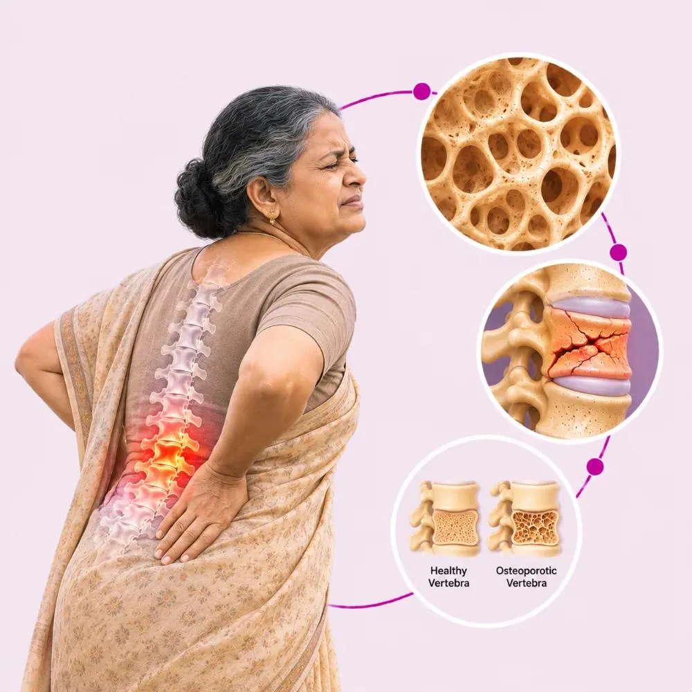
An osteoporotic spine fracture occurs when a vertebra breaks or collapses due to weakened bone strength caused by osteoporosis. Reduced bone density makes the vertebrae more vulnerable to fractures, sometimes even during normal daily activities or following minor trauma. These fractures commonly affect the thoracic (middle back) and lumbar (lower back) spine and are often called vertebral compression fractures.
Osteoporotic fractures can occur in different forms, including wedge compression fractures, where the front of the vertebra loses height; biconcave fractures, where the central portion of the vertebra becomes depressed; and crush fractures, which involve more extensive vertebral collapse.
Some patients develop fractures at a single spinal level, while others experience multiple fractures that may cause height loss, increased forward spinal curvature and persistent back pain. The fracture type, severity of symptoms and overall bone health help determine the appropriate treatment.
Reduced bone strength is the main underlying cause. Several factors can increase the risk of vertebral fractures.
Bone strength can decline with increasing age, making vertebrae more vulnerable to fracture.
Reduced oestrogen levels after menopause may accelerate bone loss and increase fracture risk.
Reduced bone mineral density is associated with a higher risk of fragility fractures.
A previous low-trauma fracture, particularly a vertebral fracture, increases the risk of further fractures.
Prolonged systemic corticosteroid use can weaken bones and contribute to osteoporosis.
Insufficient calcium intake or vitamin D deficiency may compromise bone health, depending on individual conditions.
Conditions affecting hormone balance, nutrient absorption or bone metabolism may increase fracture risk.
In people with significant osteoporosis, fractures may occur after relatively minor falls, bending, coughing or lifting.
A vertebral fracture is not always caused by a major accident. New back pain in someone at high risk of osteoporosis deserves appropriate clinical assessment.
Symptoms depend on fracture location, severity and whether surrounding nerves are affected.
Common symptoms
Emergency warning signs
Some patients experience little pain despite a vertebral fracture. Repeated fractures may contribute to progressive height loss, a rounded upper-back posture, reduced spinal mobility, chronic back discomfort, difficulty standing upright, reduced physical endurance and difficulty performing everyday activities. The emergency signs need urgent medical care: they may indicate significant nerve compression, spinal infection or another serious condition.
The diagnostic process includes determining the fracture’s age and severity and assessing underlying bone health.
The clinician reviews the onset of back pain, previous falls, height loss, medications and history of fractures. Risk factors for osteoporosis are also assessed.
The doctor examines spinal posture, tenderness and functional limitations. A neurological examination may be performed when nerve-related symptoms are present.
X-rays may show vertebral height loss, wedge deformity or other findings consistent with a compression fracture. They can help identify affected levels but may not always determine whether a fracture is recent.
MRI may be recommended when it is necessary to determine whether a fracture is recent, identify bone marrow changes or investigate possible neurological compression. It can also help distinguish certain benign fractures from fractures associated with another underlying disease.
CT provides highly detailed images of spinal bone architecture. It can help when fracture anatomy is complicated or surgical planning requires more information.
Dual-energy X-ray absorptiometry (DEXA or DXA) measures bone mineral density. A fracture-risk tool such as FRAX may also be used when appropriate to assess the likelihood of future fractures.
Additional blood tests may help identify contributing conditions such as vitamin D deficiency, kidney disease or other metabolic bone disorders. A vertebral fragility fracture is itself an important warning sign of future fracture risk, even when bone density results are not severely reduced.
Treatment has two important goals: to relieve fracture-related pain and restore safe movement, and to treat the underlying osteoporosis to reduce the risk of further fractures. Most patients begin with non-surgical management unless there is significant instability or neurological involvement.
Appropriate pain-relieving medicines may help patients remain mobile during fracture healing. Medication choice should consider age, kidney function, stomach problems, cardiovascular risk and other conditions, as older adults may be especially vulnerable to side effects from certain anti-inflammatory or sedating medicines.
Patients may need to temporarily avoid heavy lifting, repeated forward bending or activities that strongly aggravate pain. However, prolonged bed rest is generally discouraged because inactivity can worsen muscle weakness, bone loss and other complications.
A thoracolumbar brace may be considered for selected patients to improve comfort or support movement. Bracing is not routinely necessary for every compression fracture, and evidence that it improves fracture healing is limited.
Once the fracture is sufficiently stable, physiotherapy may help restore mobility, strength and confidence. A programme may include gentle mobility exercises, back extensor strengthening, balance training, posture education, walking progression, safe lifting and movement techniques, and fall-prevention exercises, adapted to fracture healing and pain tolerance.
Patients with vertebral fragility fractures often require treatment to reduce the risk of future fractures. Depending on individual risk, medicines may include bisphosphonates, denosumab or selected bone-forming therapies. The choice depends on fracture history, kidney function, bone density, other medical conditions and treatment risks.
Adequate calcium intake and vitamin D status support bone health. Supplementation may be recommended when dietary intake or clinical findings indicate a need. Do not take high-dose supplements without medical guidance.
A needle is introduced into the fractured vertebral body under imaging guidance and specialised bone cement is injected. The aim is to stabilise the treated area and potentially reduce fracture-related pain. Evidence of sustained benefit is inconsistent, so patient selection is important.
A balloon or similar device is placed inside the affected vertebra to create a cavity before bone cement is introduced. It may be considered for selected patients with severe ongoing pain from a recent, unhealed fracture despite appropriate management. Restoration of vertebral height is variable and may be limited.
Vertebral augmentation is considered only in carefully selected patients and is not recommended routinely for every osteoporotic compression fracture. A specialist may consider it when pain is severe and persistent, imaging confirms a recent, unhealed fracture at the level that matches the pain, appropriate pain management has not given sufficient relief, and the expected benefits justify the procedural risks. Vertebroplasty and kyphoplasty do not treat osteoporosis itself and must be accompanied by fracture-prevention care.
Decompression may be necessary if fracture-related changes compress neural structures.
Selected unstable fractures may require screws, rods or other devices to stabilise the spine.
Fusion may be used when long-term stabilisation of affected spinal levels is necessary.
Complex cases may require reconstruction or specialised stabilisation techniques.
Major surgery is uncommon for typical osteoporotic compression fractures. It may be considered when there is neurological compression, progressive deformity, significant instability or another structural problem. Osteoporotic bone can make surgical fixation more challenging, so careful planning is essential.
Kyphoplasty is one option for selected painful osteoporotic vertebral fractures. The actual procedure depends on the vertebral level, anatomy and clinical circumstances.
Balloon kyphoplasty
The specialist reviews symptoms, imaging, fracture age, medications and overall medical health.
Depending on the procedure and individual needs, the specialist may use general anaesthesia or local anaesthesia with sedation.
The patient is positioned to allow safe access to the affected vertebra.
Fluoroscopy or another appropriate imaging method guides instrument placement.
A specialised balloon is placed into the affected vertebral body and gently expanded to create a cavity.
Bone cement is introduced into the prepared cavity to support the treated vertebral body.
The patient is observed for pain, neurological symptoms and possible complications.
Potential risks include infection, bleeding, cement leakage, nerve irritation or injury, cement embolism, anaesthesia-related complications, persistent pain and additional vertebral fractures. Not all patients experience pain relief, and another treatment approach may be preferable.
An osteoporotic spine fracture requires more than managing immediate back pain. Clinicians must also evaluate the underlying bone weakness to reduce the risk of additional fractures.
The clinician assesses fracture location, severity and symptoms before recommending treatment.
Many stable vertebral fractures can be managed with appropriate pain relief, activity modification and rehabilitation.
Bone density testing and osteoporosis risk assessment may help guide fracture-prevention treatment.
Vertebral augmentation or surgery is reserved for the smaller number of patients for whom it is clinically necessary.
Minimising fall risk and restoring safe mobility are important components of long-term care.
Patients can contact Lux Hospitals in Hyderabad for consultations and treatment for spine fractures.
Recovery depends on fracture severity, age, general health and treatment approach. Many patients with uncomplicated vertebral compression fractures improve substantially over several weeks to approximately three months.
Pain may be significant during movement. Appropriate medication and safe mobility guidance can help reduce discomfort while avoiding unnecessary prolonged bed rest.
Some patients begin noticing gradual symptom improvement. Depending on clinical advice, patients may be encouraged to do gentle walking and basic activities.
Fracture healing continues. Rehabilitation may focus on safe movement, reducing activity-related anxiety and progressively regaining everyday function.
During this time, many stable compression fractures show significant healing and symptom improvement. Reassessment is necessary if pain persists or function deteriorates.
Patients may gradually increase physical activity and strengthening under professional guidance. Those with multiple fractures or significant deformity may require longer-term rehabilitation.
Even after their fractures heal, some people continue to have height loss or spinal curvature. Osteoporosis treatment remains important because additional fragility fractures can occur.
Avoid activities restricted by the treating clinician during healing.
Appropriate movement can help maintain physical conditioning.
Progress exercises based on fracture stability and recovery.
Review home hazards, balance, footwear, vision and medicines that may increase fall risk.
Follow prescribed osteoporosis treatment and nutritional recommendations.
Monitoring helps assess pain, function and future fracture risk.
New weakness, numbness, bladder or bowel problems, fever or rapidly worsening spinal pain require urgent assessment.
Recovery aims to restore safe movement and confidence while treating the underlying bone weakness to reduce the risk of further fractures.
Consider medical evaluation if:
Cost depends on the number of affected vertebrae, pain severity, diagnostic investigations and the treatment required. Many patients improve without surgery, while selected patients may undergo vertebral augmentation or other procedures.
| Procedure | Estimated Cost |
|---|---|
| Vertebroplasty Injects bone cement to stabilise a fractured vertebra. | ₹80,000 onward |
| Kyphoplasty Uses a balloon and bone cement to stabilise a collapsed vertebra. | ₹75,000 onward |
Fractures at more than one spinal level may change the assessment and treatment required.
Many patients improve with non-surgical management, while severe persistent pain may lead to vertebral augmentation or another procedure.
X-rays, MRI, CT scans, bone density testing and blood tests are selected according to each patient’s needs.
Non-surgical care, vertebroplasty, kyphoplasty and spinal surgery involve different equipment and clinical requirements.
Coverage depends on the insurance policy, medical necessity, exclusions and pre-authorisation requirements.
Every figure above is an indicative starting price, not a final quotation. After clinical assessment, the hospital can provide a personalised estimate based on the treatment recommended, including applicable procedure and hospital charges.

Consultant Endoscopic Spine Surgeon
MBBS, MS (Orthopaedics), Fellowship in Spine Surgery (RGUHS)
7 years experience
Common questions about osteoporotic spine fractures, healing time, bracing and kyphoplasty.
Still have questions? Call us at 079 6908 4422
Still have questions? Message us on WhatsApp at 079 6908 4444
Spine assessment at Lux Hospitals, HITEC CITY: fracture and bone health evaluation, non-surgical care first, and vertebral augmentation when it is appropriate.
Plot No.116, Lumbini Avenue, HITEC CITY, Near IKEA, Hyderabad – 500032