
Dr. Samhitha Reddy
Consultant Proctologist & Laparoscopic Surgeon
MBBS, MS, FMAS, FISCP, DMAS
- 8 Yrs
- ASCRS Member
Does walking for a few minutes cause pain, heaviness or numbness in your legs? Do your symptoms improve when you sit down or bend forward? These may be signs of spinal stenosis, a condition in which the spaces around the spinal nerves narrow.
Spinal stenosis commonly affects the lower back and neck. Some people develop significant nerve-related symptoms, while others have narrowing visible on scans without noticeable discomfort, and not every patient requires surgery. At Lux Hospitals, Hyderabad, patients can enquire about spine consultations for spinal stenosis, persistent back pain, walking difficulties and nerve-related symptoms.
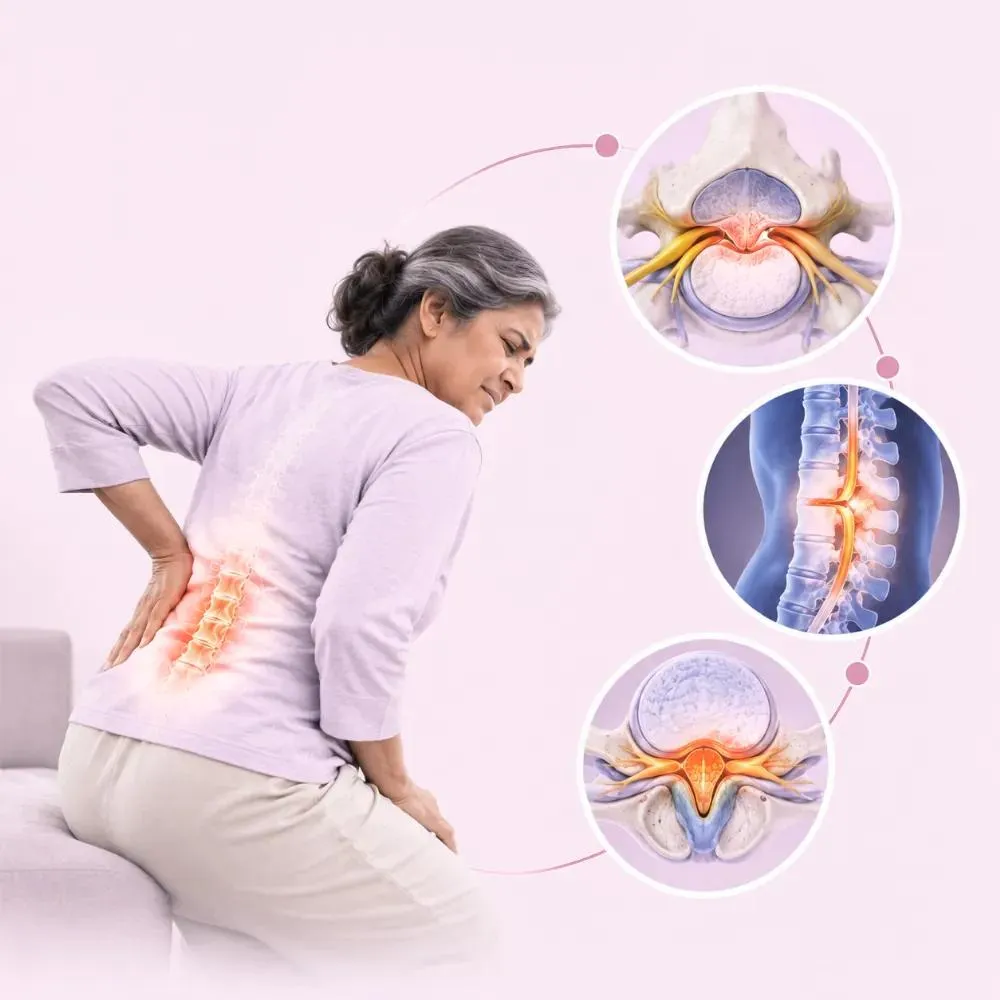
Spinal stenosis is a condition in which the spaces within the spine narrow, putting pressure on the spinal cord or nearby nerve roots. It commonly affects the lumbar spine (lower back) and cervical spine (neck). This narrowing may cause pain, numbness, tingling or muscle weakness, depending on the location and severity of nerve compression.
Lumbar spinal stenosis may cause lower back pain, leg discomfort and heaviness, particularly while standing or walking. These symptoms often improve with sitting or bending forward, a pattern known as neurogenic claudication.
Cervical spinal stenosis may cause neck pain, arm weakness, tingling or numbness. Severe narrowing can compress the spinal cord, leading to cervical myelopathy, which may affect hand coordination and balance.
Foraminal stenosis occurs when the openings through which spinal nerves exit become narrow. Treatment depends on the affected spinal region, the severity of compression and the patient’s symptoms.
Spinal stenosis most commonly develops through gradual age-related changes, although some people have a naturally narrow spinal canal.
Over time, the spine’s discs, joints and ligaments undergo degenerative changes. These changes may gradually reduce the space available for nerve structures.
Arthritis affecting the spine’s small joints may lead to bony growths called osteophytes, or bone spurs. These can narrow the spinal canal or the openings where nerves exit.
Ligaments supporting the spine may become thicker with age. The ligamentum flavum, located towards the back of the spinal canal, can contribute to nerve compression when it thickens.
Degenerative disc bulges and herniations can reduce the available space around spinal nerves.
Spondylolisthesis occurs when one vertebra slips forwards or backwards on another. In some patients, this contributes to spinal narrowing and nerve compression.
Some people are born with a narrower-than-usual spinal canal and may experience symptoms earlier in life.
Trauma, certain spinal conditions and postoperative changes may occasionally contribute to narrowing.
Age-related degeneration is common, but a scan showing spinal narrowing does not always mean the condition is causing symptoms.
Symptoms vary according to the location and severity of narrowing.
Common symptoms of lumbar spinal stenosis
Emergency warning signs
Some patients find walking while leaning forward, such as when pushing a shopping trolley, more comfortable than walking upright. Cervical spinal stenosis may instead cause neck discomfort, pain spreading into the shoulder or arm, numbness or tingling in the hands, reduced hand strength, difficulty with fine hand movements, balance problems and walking difficulties. The emergency signs can indicate severe nerve or spinal cord compression and need immediate emergency medical attention. Early assessment is particularly important for suspected cervical myelopathy, because spinal cord dysfunction can progress.
Diagnosis involves a detailed medical history, physical examination and appropriate imaging.
The doctor reviews pain location, symptom duration and changes associated with walking, standing or sitting. The symptom pattern helps distinguish spinal stenosis from other causes of back or leg pain.
The clinician may examine muscle strength, sensation, reflexes and walking ability. For cervical stenosis, hand coordination, balance and signs of spinal cord involvement may also be evaluated.
X-rays may help identify degenerative changes, spinal alignment problems, spondylolisthesis or other bony abnormalities. However, they do not show the spinal cord and nerve roots in detail.
An MRI can show the spinal cord, nerve roots, discs, ligaments and spinal canal in great detail. It is frequently used when symptomatic spinal stenosis needs further investigation or surgical planning.
CT may provide more information about bone structures when MRI is not suitable or when greater anatomical detail is needed.
The clinician evaluates walking tolerance, neurological findings and how symptoms affect daily activities.
Imaging findings are interpreted alongside the examination before deciding between conservative care and surgery.
Treatment depends on symptom severity, neurological findings, the affected spinal level and overall health. Many patients with lumbar spinal stenosis can control their symptoms without surgery. However, specialist evaluation is necessary if there is substantial neurological decline or progressive spinal cord compression.
May help improve muscle strength, movement and walking tolerance. A programme may include gentle mobility exercises, core and trunk strengthening, hip and leg conditioning, balance exercises when appropriate, gradual walking or cycling programmes and functional movement training, adapted to symptoms and neurological findings.
Certain activities, particularly prolonged upright standing or walking, may aggravate lumbar stenosis symptoms. Taking breaks, adjusting posture and using appropriate mobility support can help some patients.
Selected pain-relieving or anti-inflammatory medicines may be considered after reviewing risks involving the stomach, kidneys and cardiovascular system. Medication should be individualised.
Structured rehabilitation can help maintain physical conditioning and improve confidence during movement. Although exercise does not physically widen the narrowed spinal canal, it may improve symptoms and function.
Injections are sometimes considered for selected nerve-related symptoms, but evidence for sustained benefit in central lumbar spinal stenosis is limited. They are not a routine solution for neurogenic claudication: NICE specifically advises against epidural steroid injections for neurogenic claudication caused by central spinal canal stenosis.
When appropriate, weight control, stopping smoking and regular, moderate physical activity can support overall spinal and cardiovascular health.
Removes a portion of bone and associated structures that contribute to spinal canal narrowing. The aim is to provide more space for compressed nerve structures.
Removes a small portion of the vertebral lamina to relieve nerve pressure. It may be suitable for selected cases.
Widens a narrowed opening through which a spinal nerve exits. It may be used when foraminal stenosis compresses a nerve.
Selected decompression procedures may be performed through smaller approaches using specialised instruments. Suitability depends on spinal anatomy, the extent of narrowing and surgical expertise.
Cervical stenosis causing spinal cord compression may require procedures such as cervical laminectomy, laminoplasty or anterior cervical decompression. The choice depends on the location of compression, spinal alignment and other clinical factors.
May be considered when spinal instability, deformity or other specific conditions require stabilisation. It is not automatically required for every spinal stenosis operation.
Surgery may be advised when serious symptoms persist despite appropriate conservative care, or when neurological impairment requires intervention.
When lumbar spinal stenosis causes significant nerve compression, decompression surgery may be recommended. A lumbar laminectomy is one established approach.
Lumbar laminectomy
The operation is commonly performed under general anaesthesia. The anaesthesia team monitors the patient’s vital functions throughout the procedure.
The patient is positioned to allow appropriate access to the affected spinal area.
The surgeon makes an incision over the relevant spinal level. The approach may be open or minimally invasive, depending on the clinical situation.
The surgeon locates the structures causing compression, such as thickened ligaments or bone overgrowth.
The surgeon removes the necessary bone or ligament tissue to create more space for the affected nerves.
The surgeon decides whether decompression alone is adequate or whether stabilisation is required. Fusion is performed only when clinically required.
The surgeon closes the wound and the patient is observed while recovering from anaesthesia. Early mobility may be encouraged when medically appropriate.
The approach chosen for you depends on the location and extent of narrowing, your anatomy and your symptoms.
Appropriate spinal stenosis care starts with determining whether spinal narrowing is causing the patient’s symptoms and whether there is significant nerve or spinal cord involvement.
Symptoms, examination findings and relevant imaging guide treatment decisions.
Patients without progressive neurological impairment may benefit from appropriate rehabilitation and conservative management.
Increasing weakness, walking difficulties and changes in coordination require careful assessment.
Lumbar stenosis, cervical stenosis and foraminal narrowing may require different approaches.
Lux Hospitals is located in HITEC CITY, near IKEA, with access from Gachibowli and surrounding areas of Hyderabad.
Recovery varies depending on whether the patient receives non-surgical care, decompression surgery or decompression with fusion.
Patients beginning conservative care may focus on symptom management, appropriate movement and avoiding prolonged inactivity. After surgery, monitoring and early assisted mobility may begin when medically appropriate.
Some patients who have had decompression surgery may begin returning to light daily activities. Wound care, walking and activity restrictions should follow the surgical team’s instructions.
Patients may gradually improve walking tolerance and physical function. Rehabilitation may focus on mobility, strengthening and balance.
Some patients progress towards increased activity and work responsibilities. Recovery may be slower after more extensive operations or fusion.
Continued rehabilitation may help improve strength and endurance. Patients with longstanding neurological symptoms may need more time and may not fully recover.
Exercise progression should reflect the procedure, pain level and neurological condition.
Adjust walking distance based on tolerance and medical advice.
Heavy lifting and strenuous activity may need temporary restrictions after surgery.
A balanced diet, exercise and treatment of long-term illnesses may support recovery.
Postoperative reviews help monitor wound healing, neurological symptoms and functional progress.
Get checked immediately if you develop severe new symptoms, such as increasing weakness, fever, wound problems or new bowel or bladder problems.
Recovery is measured by neurological function and progress, not just by whether pain eases straight away.
Consider a spine consultation if:
The cost of spinal stenosis treatment in Hyderabad depends on the affected spinal level, the degree of nerve compression, the treatment approach and medical requirements. Not every patient requires surgery: some manage their symptoms with physiotherapy, medicines and activity modification.
| Procedure | Estimated Cost |
|---|---|
| Laminectomy (Lumbar) Removes bone or tissue to relieve spinal nerve compression. | ₹95,000 onward |
| Endoscopic Spine Surgery Relieves nerve compression using minimally invasive techniques. | ₹1,40,000 onward |
| Interspinous Spacer Places an implant between vertebrae to reduce nerve compression in selected patients. | ₹1,70,000 onward (estimate) |
| Spinal Fusion Stabilises affected vertebrae when spinal instability is present. | ₹1,89,000 onward |
Where the narrowing lies, and how much of the spine is involved, influences the treatment recommended.
The severity of compression helps decide between conservative care and surgery.
Physiotherapy, medicines and activity modification involve different requirements from surgical treatment.
Surgical costs depend on whether decompression alone is required or additional stabilisation procedures are necessary.
Every figure above is an indicative starting price, not a final quotation. The initial consultation may include a clinical examination, a neurological evaluation and a review of previous imaging, after which the hospital can provide a personalised estimate based on the treatment recommended.

Consultant Endoscopic Spine Surgeon
MBBS, MS (Orthopaedics), Fellowship in Spine Surgery (RGUHS)
7 years experience
Common questions about spinal stenosis, walking, recovery and when surgery is considered.
Still have questions? Call us at 079 6908 4422
Still have questions? Message us on WhatsApp at 079 6908 4444
Spine assessment at Lux Hospitals, HITEC CITY: a clear diagnosis, non-surgical options first, and decompression surgery when it is needed.
Plot No.116, Lumbini Avenue, HITEC CITY, Near IKEA, Hyderabad – 500032