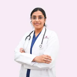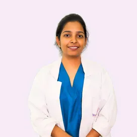
Dr. Samhitha Reddy
Consultant Proctologist & Laparoscopic Surgeon
MBBS, MS, FMAS, FISCP, DMAS
- 8 Yrs
- ASCRS Member
Most growths on the skin are entirely harmless: moles, skin tags, cysts and warts that have been there for years. A small number are not, and the ones that matter are usually distinguishable by how they have changed rather than by how they look on any single day.
A lesion that is new, growing, changing colour, bleeding or refusing to heal deserves a proper look. Our surgeons at Lux Hospitals, HITEC CITY assess, biopsy and excise skin tumours with clear margins, and every specimen goes for histopathology.
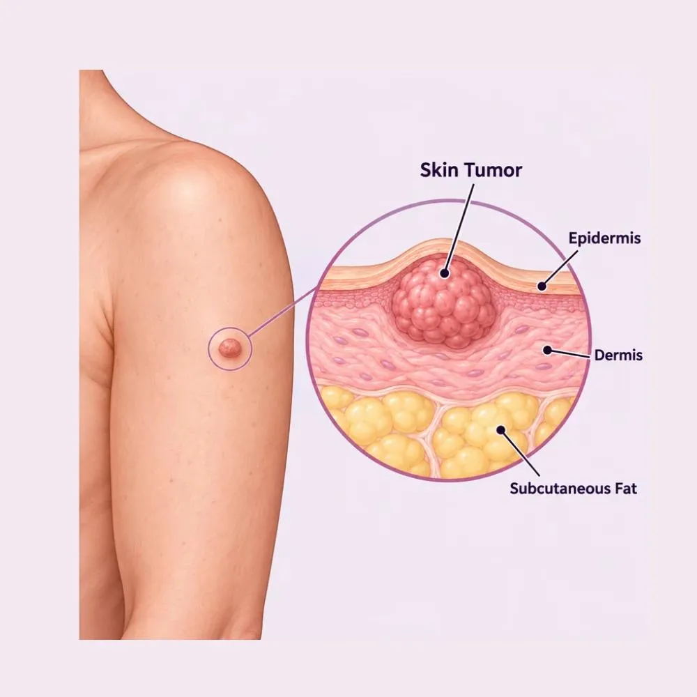
A skin tumour is any abnormal growth of cells in the skin. The great majority are benign (moles, seborrhoeic keratoses, lipomas, sebaceous cysts, skin tags and dermatofibromas) and cause no harm beyond appearance or irritation.
The malignant ones fall into three main types. Basal cell carcinoma is the commonest, grows slowly, and very rarely spreads, but it does erode locally if left. Squamous cell carcinoma grows faster and can spread to lymph nodes. Melanoma is the least common of the three and the most serious, because it can spread early, which is exactly why it is worth catching early.
The distinction between these cannot be made reliably by eye, even by an experienced surgeon. That is why anything suspicious is biopsied or excised and examined under a microscope, rather than watched and hoped about.
Ultraviolet exposure is the dominant cause of skin cancer, and it is the one factor almost entirely within your control.
Cumulative ultraviolet exposure damages DNA in skin cells. It is the single largest risk factor for all three types of skin cancer.
Episodes of severe blistering sunburn, particularly in childhood, substantially increase melanoma risk decades later.
A first-degree relative with melanoma raises your own risk considerably, and warrants a lower threshold for having lesions checked.
Lighter skin types burn more readily, and having more than fifty moles is an independent risk factor for melanoma.
Organ transplant recipients, people on long-term immunosuppression and those with HIV develop skin cancers considerably more often.
Arsenic, industrial tars and certain occupational chemicals are established causes of squamous cell carcinoma.
Skin within a previously irradiated field carries a raised long-term risk of developing tumours.
Squamous cell carcinoma can arise in long-standing ulcers, burns scars and chronic inflammatory skin conditions.
Sun protection genuinely works, and it works at any age. Shade in the middle of the day, covering up, and daily sunscreen reduce the risk of every type of skin cancer.
Change is the thing to watch for. A lesion that has looked the same for twenty years is far less concerning than one that has altered over three months.
Common benign features
Warning signs: the ABCDE rule
A lesion that bleeds without being knocked, itches persistently, or fails to heal is worth showing someone. So is a mole that looks noticeably different from all your others.
Examination narrows the possibilities; only histopathology settles the question. Anything suspicious is sampled rather than monitored.
The lesion is assessed for size, colour, border, texture and fixation, along with a check of the surrounding skin and nearby lymph nodes.
A magnified, illuminated view of the lesion revealing pigment patterns and vessel structures invisible to the naked eye.
A small sample taken under local anaesthetic where the lesion is large or in a site where full excision would be a major undertaking.
The whole lesion is removed with a narrow margin. Preferred for suspected melanoma, since it lets the pathologist measure the full depth.
Assesses the draining lymph nodes where a squamous cell carcinoma or melanoma has been confirmed.
CT or PET-CT for confirmed melanoma or advanced squamous cell carcinoma, to check whether it has spread beyond the skin.
For suspected melanoma, the lesion is removed whole rather than sampled. The pathologist measures how deep it goes, and that measurement determines everything that follows.
Treatment depends entirely on the type, size, depth and location of the tumour. Benign lesions need removal only if they are troublesome; malignant ones need removal with an adequate margin.
Liquid nitrogen freezes and destroys abnormal cells. Effective for small superficial lesions, actinic keratoses and warts, done in the clinic in minutes.
Creams containing 5-fluorouracil or imiquimod applied over weeks, for superficial basal cell carcinoma and pre-cancerous patches.
Focused light removes or destroys superficial lesions with precision and minimal scarring, suited to cosmetically sensitive areas.
The lesion is scraped away and the base cauterised. Quick and effective for small, well-defined, low-risk basal cell carcinomas.
For tumours where surgery is unsuitable (difficult sites, elderly or frail patients) or as additional treatment after excision.
Benign lesions that are stable, asymptomatic and confidently diagnosed need no treatment at all.
The standard treatment. The tumour is removed with a margin of healthy tissue, the width of which is set by the tumour type and thickness.
A larger margin for melanoma and higher-risk squamous cell carcinoma, sized according to how deep the tumour has grown.
The tumour is removed layer by layer, each checked under the microscope during the procedure until margins are clear. Spares the most healthy tissue, ideal on the face.
The first draining lymph node is identified and removed to check for spread, in melanoma above a certain thickness.
Skin grafts or local flaps to close a defect too large to stitch directly, restoring both function and appearance.
The single most important thing in treating a malignant skin tumour is clear margins confirmed by the pathologist. Everything else (the technique, the closure, the cosmetic result) is arranged around achieving that.
A twenty to sixty minute day-care procedure depending on size and site. You are awake, the area is completely numb, and you go home the same day.
Surgical excision under local anaesthetic
The lesion is examined, often with dermoscopy, and the excision margin is marked on the skin: its width determined by the suspected diagnosis.
Local anaesthetic is injected around the lesion. A brief sting, after which you feel pressure but no pain.
The lesion is removed as an ellipse of skin including the planned margin, taken down to an appropriate depth for the tumour type.
The specimen is marked with a suture so the pathologist can report exactly which edge is involved if a margin is not clear. This makes any further surgery precise.
Closed directly with sutures where possible. Larger defects are closed with a local flap or a skin graft, planned to follow natural skin lines.
A dressing is applied and you go home within the hour, with wound care instructions and a date to receive the histopathology result.
The result confirms the diagnosis and whether margins are clear. Where a margin is involved, a further wider excision is arranged.
The histopathology appointment is the important one. It gives you the definitive diagnosis, confirms the tumour was fully removed, and sets what follow-up, if any, you need.
Skin tumour surgery is judged on two things: whether the margins were clear, and how the scar looks. Here is why patients trust us:
Surgeons experienced in excising benign and malignant skin lesions, including reconstruction where a defect cannot be closed directly.
Dermoscopy, biopsy and histopathology on every specimen, with oriented specimens so any further surgery is precisely targeted.
From the first consultation through to post-treatment follow-up, with one team throughout.
Our hospital is in HITEC CITY, near IKEA: easily reachable from across Hyderabad.
Recovery after a simple excision is quick. Where a graft or flap was used it takes longer, and the dressing regime is more involved.
Home within the hour. Mild soreness as the anaesthetic wears off, controlled with simple painkillers.
Keep the dressing clean and dry. Normal daily activity resumes, avoiding stretching or straining the wound.
Sutures removed, timing depending on the site: around five days on the face, ten to fourteen on the trunk or limb.
The wound is healed. Histopathology is discussed and any further treatment planned. Full activity resumes.
The scar softens and fades progressively. Ongoing skin surveillance where a malignancy was confirmed.
Cover it or use high-factor sunscreen for at least six months. This matters doubly here: sun caused the problem in the first place.
Follow the dressing instructions exactly, particularly with a skin graft, where the first dressing is left undisturbed.
Having had one skin cancer raises the chance of another. A monthly self-check, including your back with a mirror, catches new lesions early.
No heavy lifting or vigorous exercise until sutures are out, particularly for wounds over a joint or on the back.
Fever, spreading redness, increasing pain, discharge, wound edges separating, or a graft turning dark.
The pathology appointment is essential. Where a malignancy was confirmed, regular skin surveillance follows for several years.
Sun protection is the aftercare that matters most. Daily sunscreen, shade in the middle of the day, and covering up reduce the risk of the next one considerably.
Book a consultation if any of the following apply.
Cost depends on the type and extent of the tumour, the technique used, and whether reconstruction is needed. These are the ranges our patients fall into.
| Procedure | Estimated Cost |
|---|---|
| Consultation & dermoscopy Per visit | ₹800 – ₹2,000 |
| Cryotherapy Per session, for small superficial lesions | ₹2,000 – ₹6,000 |
| Punch biopsy Including histopathology | ₹4,000 – ₹9,000 |
| Excision of a benign lesion Day care, including histopathology | ₹15,000 – ₹35,000 |
| Excision of a malignant tumour Day care, with margin assessment | ₹40,000 – ₹90,000 |
| Wide excision with flap or graft Day care or one night’s stay | ₹80,000 – ₹1,80,000 |
| Excision with sentinel node biopsy One to two nights’ stay | ₹1,50,000 – ₹2,80,000 |
A benign mole excised for cosmetic reasons is straightforward. A melanoma needs a wide margin, node assessment and staging.
A lesion on the back can usually be closed directly. The same lesion on the nose or eyelid may need a flap or graft.
A defect too large to close directly requires a graft or local flap, which adds significantly to theatre time.
A shared ward, a twin-sharing room or a private suite: the largest single swing in most estimates.
We are empanelled with most major insurers, and our team files the pre-authorisation for you.
Every figure above is an indicative range, not a quote. After your consultation you are given a written, fixed-cost estimate, and that is the number you pay, with no additions once treatment begins.
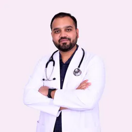
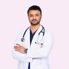
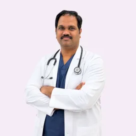
The general surgeons who diagnose and excise skin tumours at our HITEC CITY hospital.
Consultant General & Advanced Laparoscopic Surgeon
MBBS, MS, FMAS
17 years experience

Senior Consultant: Bariatric & Gastrointestinal Surgery
MBBS, MS, FRCS (UK), DLS (France)
28 years experience

Consultant Proctologist & Laparoscopic Surgeon
MBBS, MS, FMAS, FISCP, DMAS
8 years experience
Reviews from patients treated for skin lesions, lipoma and general surgery at Lux Hospitals, Hyderabad.
Written by Dr. Abhishek Katha on skin lesions, lumps and general surgical care.

Dr. Abhishek Katha
Dr. Abhishek Katha

Dr. Abhishek Katha

Dr. Abhishek Katha
Dr. Abhishek Katha

Dr. Abhishek Katha
Common questions about skin tumours, their removal, and what treatment costs at Lux Hospitals.
Still have questions? Call us at 079 6908 4422
Still have questions? Message us on WhatsApp at 079 6908 4444
Accurate diagnosis and clear-margin excision of skin tumours, with a fixed-cost estimate before treatment begins.
Plot No.116, Lumbini Avenue, HITEC CITY, Near IKEA, Hyderabad – 500032