
Dr. Samhitha Reddy
Consultant Proctologist & Laparoscopic Surgeon
MBBS, MS, FMAS, FISCP, DMAS
- 8 Yrs
- ASCRS Member
Blood in the urine is a symptom, not a diagnosis. It always has a cause, and finding that cause is the entire point of the consultation.
Most causes are benign: a stone, an infection, an enlarged prostate. But painless visible blood is the classic first sign of a bladder or kidney tumour, and it is the one symptom in urology that is never dismissed. Our urologists at Lux Hospitals, HITEC CITY investigate it fully, even when it happens once and stops.
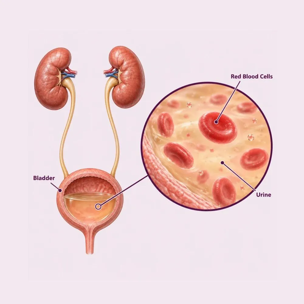
Hematuria means blood in the urine. It comes in two forms. Gross or visible hematuria is blood you can see: the urine appears pink, red, or the colour of tea or cola. Microscopic hematuria is invisible to the eye and detected only on a urine test, often done for something else entirely.
Both need investigating, but visible hematuria carries considerably more weight. Around one in five people with painless visible blood in the urine turns out to have a urological cancer, most commonly of the bladder. For microscopic hematuria the figure is far lower, but not zero, which is why it too is assessed rather than ignored.
The timing during the stream is genuinely informative. Blood at the very start of urination suggests a urethral source. Blood throughout the stream points to the bladder, ureter or kidney. Blood only at the end suggests the bladder neck or prostate. It is a simple observation that meaningfully narrows the search before any test is done.
Causes range from entirely benign to serious, and they cannot be told apart without investigation.
The commonest cause, particularly in women. Blood comes with burning and frequency, and settles once the infection is treated.
A stone abrading the lining of the kidney, ureter or bladder. Typically accompanied by severe colicky pain.
The reason all hematuria is investigated. Bladder cancer classically causes painless visible blood, often intermittently, and smoking is the dominant risk factor.
An enlarged prostate has fragile surface vessels that bleed, particularly at the end of the stream. A common cause in men over fifty.
Glomerulonephritis and IgA nephropathy cause microscopic hematuria alongside protein in the urine, often with tea-coloured urine after an infection.
Strenuous exercise, particularly running, can cause transient hematuria. Direct injury to the kidney or bladder causes it more obviously.
Anticoagulants make bleeding more visible but do not cause it. Hematuria on a blood thinner still needs full investigation: the drug unmasked a cause rather than creating one.
Beetroot, some medications and certain food dyes can turn urine red without any blood present. A urine test distinguishes them in minutes, which is why the test comes before the worry.
What accompanies the blood, and when in the stream it appears, points strongly toward the cause.
How it presents
Features needing urgent care
Painless visible blood is the pattern that concerns urologists most, precisely because it is painless. There is nothing to prompt urgency, so people wait, and it is the presentation most strongly associated with bladder cancer.
The workup covers the whole urinary tract, because blood can originate anywhere along it.
Confirms genuine blood rather than dye or pigment, detects infection, and looks for protein and casts suggesting kidney disease.
Examines urine for abnormal cells shed from a bladder tumour. Useful when positive, though it does not exclude cancer when negative.
Creatinine and eGFR for kidney function, haemoglobin to assess blood loss, clotting studies, and PSA in men.
The standard imaging for visible hematuria. It images kidneys, ureters and bladder in detail and detects stones and tumours ultrasound would miss.
A camera passed into the bladder. The only way to see the bladder lining directly, and essential: imaging alone misses small flat tumours.
Where microscopic hematuria comes with protein loss and reduced function, pointing to intrinsic kidney disease rather than a urological cause.
CT and cystoscopy are complementary rather than alternatives. CT sees the kidneys and ureters that the scope cannot reach; the scope sees flat bladder lesions that no scan detects. Visible hematuria warrants both.
Hematuria itself is not treated: the cause is. What follows depends entirely on what the investigation finds.
Culture-guided treatment, with a repeat urine test afterwards to confirm the blood has cleared. Persistent blood after treatment needs further investigation.
Hydration, pain relief and medical expulsive therapy for small stones, with endoscopic treatment for larger ones.
Alpha blockers and 5-alpha reductase inhibitors, which reduce prostate bleeding as well as obstruction.
Blood pressure control and specific treatment for glomerulonephritis, in conjunction with kidney specialists.
Adjusting blood thinners where bleeding is heavy, in discussion with the prescribing doctor: never stopped unilaterally.
Transurethral resection of a bladder tumour through the urethra, which both removes it and provides tissue for staging. No incision.
Ureteroscopy with laser, or PCNL for larger stones, removing the source of bleeding definitively.
Urgent clearance of clots causing retention, through a wide catheter, with continuous irrigation until bleeding settles.
TURP or laser prostatectomy where an enlarged prostate is the confirmed source of recurrent bleeding.
Removal of a kidney tumour, preserving as much kidney as the tumour allows.
Clot retention, where clots block the bladder outlet completely, is a urological emergency. It needs a wide catheter and washout the same day, and is one of the few presentations that cannot wait.
Visible hematuria follows a defined pathway, designed so that nothing along the tract is missed.
Structured investigation of the whole urinary tract
A urine test distinguishes actual blood from beetroot, dyes and medication. This takes minutes and settles a good number of referrals immediately.
Timing during the stream, associated pain, smoking history, occupational exposure, medications and previous episodes.
Urine culture, with infection treated first. If blood persists after successful treatment, the workup continues regardless.
Kidney function, haemoglobin, clotting and PSA, alongside urine cytology for abnormal cells.
Imaging the kidneys, ureters and bladder with contrast, detecting stones, masses and abnormalities of the collecting system.
Direct inspection of the bladder lining, usually under local anaesthesia as a day-care procedure, with biopsy of anything abnormal.
Definitive treatment of whatever is found, with follow-up urine testing to confirm the bleeding has resolved.
A negative workup is a genuinely good result, not a wasted one. It means the serious causes have been excluded, and it is worth having on record if bleeding recurs later.
Blood in the urine needs a complete answer, not a partial one. Here is why patients trust us:
CT urogram and cystoscopy together where visible hematuria is confirmed, because each detects what the other misses.
Flexible cystoscopy as a day-care procedure under local anaesthesia, without referral or delay.
From the first consultation through to post-treatment follow-up, with one team throughout.
Our hospital is in HITEC CITY, near IKEA: easily reachable from across Hyderabad.
The investigation itself is straightforward. What follows depends entirely on the diagnosis.
Urine and blood tests, ultrasound, and treatment of any infection found. Many cases are resolved at this point.
CT urogram and cystoscopy where visible hematuria is confirmed. Cystoscopy is day care with mild burning for a day or two afterwards.
Results reviewed together. In the majority of cases this is a reassuring conversation with no further action needed.
Stone or tumour treatment planned, generally endoscopic, with a short admission and one to two weeks off work.
Repeat urine testing to confirm clearance. After a bladder tumour, regular surveillance cystoscopy for several years.
Burning and slight blood for a day or two is normal. High fluid intake flushes the tract and shortens it.
Smoking is the dominant risk factor for bladder cancer. If your hematuria workup was negative, this is the single most valuable thing to take from it.
Blood returning after a negative workup needs reassessment. Bladder tumours can bleed intermittently and a single clear result does not close the matter permanently.
After a bladder tumour, repeat cystoscopy detects recurrence early when it remains easily treatable. This is essential rather than optional.
Hematuria on anticoagulation still needs investigating, but stopping the drug without advice risks stroke or clot. Discuss it rather than deciding alone.
Clots in the urine, or an inability to pass urine, need same-day treatment with a catheter and bladder washout.
Blood that appears once and stops still needs investigating. Intermittent bleeding is characteristic of bladder tumours, and treating a single episode as resolved is the commonest way a diagnosis is delayed.
Book a consultation if any of the following apply.
The investigation is the main cost. Treatment depends entirely on what is found, and many people need none.
| Procedure | Estimated Cost |
|---|---|
| Consultation Includes examination | ₹800 – ₹1,500 |
| Urine tests and culture Analysis, culture and cytology | ₹800 – ₹2,500 |
| Blood workup Kidney function, haemoglobin, clotting, PSA | ₹1,500 – ₹4,000 |
| Ultrasound KUB First-line imaging | ₹1,500 – ₹3,500 |
| CT urogram Standard for visible hematuria | ₹6,000 – ₹14,000 |
| Flexible cystoscopy Day care, local anaesthesia | ₹12,000 – ₹25,000 |
| TURBT for bladder tumour Two to three days in hospital | ₹1,20,000 – ₹2,40,000 |
Visible hematuria needs the full pathway including CT and cystoscopy. Microscopic hematuria in a low-risk patient may need considerably less.
An infection is treated with a course of antibiotics. A stone or tumour needs a procedure, which is the substantial cost.
Clot retention needs emergency admission, catheterisation and washout, which costs more than an elective outpatient pathway.
A shared ward, a twin-sharing room or a private suite: the largest single swing in most estimates.
We are empanelled with most major insurers, and our team files the pre-authorisation for you.
Every figure above is an indicative range, not a quote. After your consultation you are given a written, fixed-cost estimate, and that is the number you pay, with no additions once treatment begins.
The urologists who investigate blood in the urine at our HITEC CITY hospital.

Consultant Urologist & Andrologist
MBBS, MS, DrNB (Urology)
10 years experience

Consultant Urologist & Andrologist
MBBS, MS, MCh (Urology)
9 years experience
Reviews from patients treated for kidney and urological conditions at Lux Hospitals, Hyderabad.
Written by Dr. Vipin Reddy B on kidney health and urological care.

Dr. Vipin Reddy B

Dr. Vipin Reddy B

Dr. Vipin Reddy B

Dr. Vipin Reddy B

Dr. Vipin Reddy B

Dr. Vipin Reddy B
Common questions about blood in the urine, how it is investigated and what it costs at Lux Hospitals.
Still have questions? Call us at 079 6908 4422
Still have questions? Message us on WhatsApp at 079 6908 4444
Complete investigation of blood in the urine, including CT urogram and cystoscopy, with costs shared upfront.
Plot No.116, Lumbini Avenue, HITEC CITY, Near IKEA, Hyderabad – 500032