
Dr. Samhitha Reddy
Consultant Proctologist & Laparoscopic Surgeon
MBBS, MS, FMAS, FISCP, DMAS
- 8 Yrs
- ASCRS Member
The kidney, ureter and bladder form one continuous system, and a problem in any part of it affects the rest. Urologists refer to it as the KUB, and it is assessed as a whole rather than piece by piece.
A stone in the ureter causes back-pressure on the kidney. An enlarged prostate blocking the bladder outlet eventually damages the kidneys above it. Our urologists at Lux Hospitals, HITEC CITY investigate the whole tract, find where the problem actually lies, and treat the cause rather than the symptom.
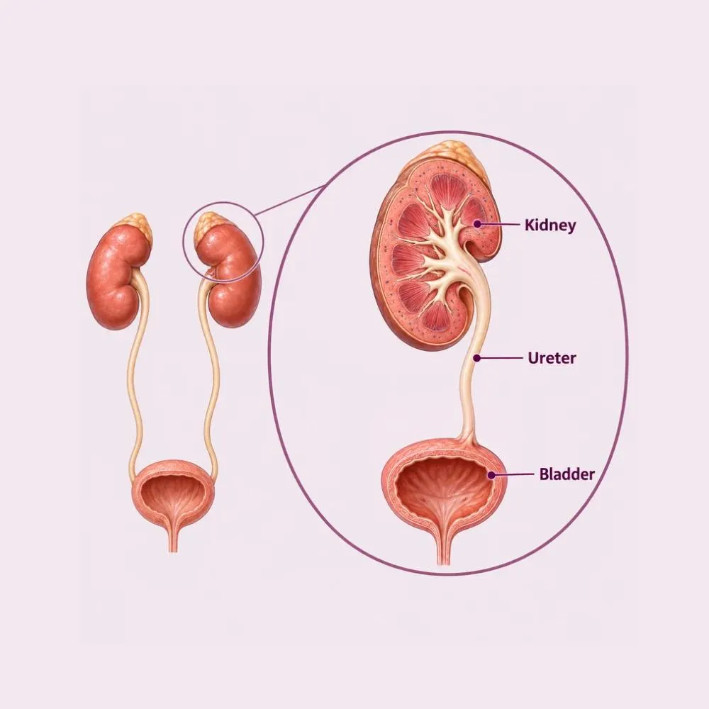
The urinary tract begins at the kidneys, which filter blood and produce urine. Urine travels down two muscular tubes called ureters into the bladder, which stores it, and leaves through the urethra. Each part has a distinct job, and problems in each produce distinct symptoms, which is why the pattern of what you experience points to where the trouble is.
The reason urologists assess the whole tract rather than one part is back-pressure. Urine flows in one direction, and anything obstructing that flow raises pressure upstream. A stone lodged in the ureter causes the kidney above it to swell. An enlarged prostate obstructing the bladder outlet leads, over years, to a thickened bladder and eventually to damaged kidneys. Treating only what hurts misses the part that is quietly failing.
KUB conditions span stones, infections, obstruction, incontinence, cancers and congenital abnormalities. The investigations are largely common to all of them (a urine test, a blood creatinine, an ultrasound and often a CT) and it is the combination that localises the problem.
A handful of conditions account for the large majority of urological presentations.
Crystals forming in concentrated urine, which can lodge anywhere from the kidney to the urethra. Particularly common in Hyderabad’s climate.
Bacteria ascending from the urethra into the bladder and occasionally the kidney. Far commoner in women because of a shorter urethra.
Benign prostatic hyperplasia obstructing the bladder outlet in men over fifty. Untreated, it eventually affects the bladder and kidneys.
Narrowing of the ureter or urethra from scarring, previous instrumentation or congenital abnormality, causing back-pressure.
Overactive bladder, neurogenic bladder from diabetes or spinal disease, and pelvic floor weakness after childbirth.
Kidney, ureteric, bladder and prostate cancers. Painless visible blood in the urine is the classic presentation and always needs investigating.
Trauma, and abnormalities such as reflux or pelviureteric junction obstruction present from birth.
Painless blood in the urine is the symptom never to dismiss. It is often the first sign of a bladder or kidney tumour, and it is investigated fully even when it happens once and stops.
The site of the symptom usually indicates the site of the problem, which is what makes the history so informative.
Kidney and ureter symptoms
Bladder and outflow symptoms
Fever with flank pain, complete inability to pass urine, or visible blood in the urine are all reasons to be seen the same day rather than to wait for an appointment.
A common set of investigations covers most of the tract, and the combination localises the problem.
Detects infection, blood, protein and crystals, and identifies the organism and the antibiotic it responds to.
Creatinine and eGFR for kidney function, plus PSA in men where prostate disease is suspected.
Images kidneys, bladder and post-void residual volume. Non-invasive, no radiation, and the first-line test for most presentations.
The definitive test for stones and for the ureters, which ultrasound visualises poorly. It maps the entire tract in detail.
A camera passed into the bladder through the urethra. The only way to see the bladder lining directly, and essential in unexplained haematuria.
Measures bladder pressure and flow during filling and voiding, distinguishing obstruction from a bladder that is not contracting properly.
Ultrasound sees the kidneys and bladder well but the ureters poorly, since they lie behind bowel gas. That is why a CT urogram is added whenever a ureteric problem is suspected.
Most urinary tract conditions are treated medically or endoscopically. Open surgery is now uncommon.
Guided by urine culture rather than given blindly, with longer courses for kidney infection than for simple cystitis.
Alpha blockers to help a small ureteric stone pass, alongside good hydration and pain relief.
Alpha blockers and 5-alpha reductase inhibitors for prostate obstruction; anticholinergics or beta-3 agonists for overactive bladder.
Structured exercises and bladder training for stress and urge incontinence, effective in a substantial proportion of cases.
High fluid intake to prevent stones, with dietary modification guided by stone composition where available.
A fine scope passed up the ureter to fragment and remove a stone with a laser. No incision at all.
Percutaneous nephrolithotomy through a small tract into the kidney, for large or staghorn stones.
Endoscopic removal of obstructing prostate tissue through the urethra, relieving outlet obstruction definitively.
Endoscopic resection of a bladder tumour, which both treats the tumour and provides tissue for staging.
Urgent drainage of an obstructed kidney, relieving back-pressure and infection while definitive treatment is planned.
Urethroplasty for stricture, pyeloplasty for pelviureteric obstruction, and bladder reconstruction in selected severe cases.
An obstructed, infected kidney is one of the true urological emergencies. Draining it with a stent or nephrostomy takes priority over treating the underlying stone, which is dealt with once the infection has settled.
The pathway is the same regardless of the eventual diagnosis: localise the problem, then treat the cause.
Structured urological pathway
The pattern and site of symptoms, urinary habits, previous stones or infections, and an abdominal and genital examination.
Urinalysis and culture, creatinine and eGFR, and PSA where relevant. These often narrow the diagnosis substantially on their own.
Assessing kidney size, hydronephrosis, bladder wall thickness and residual volume after voiding.
CT urogram where stones or ureteric disease are suspected, and cystoscopy where the bladder lining needs direct inspection.
Urgent catheter, stent or nephrostomy where the tract is blocked, particularly with infection or impaired function.
Medical, endoscopic or surgical treatment of the underlying cause, planned once the picture is complete and the patient is stable.
Repeat imaging and function tests, plus preventive measures such as fluid targets after stones or surveillance after bladder tumours.
Relieving obstruction comes before everything else. A blocked kidney loses function measurably within days, and that loss does not fully return.
The urinary tract is one system, and treating it in parts leads to missed diagnoses. Here is why patients trust us:
Kidneys, ureters, bladder and outflow investigated together, so back-pressure and silent kidney damage are found rather than missed.
Laser, ureteroscopic and transurethral techniques wherever possible, so open surgery is genuinely a last resort.
From the first consultation through to post-treatment follow-up, with one team throughout.
Our hospital is in HITEC CITY, near IKEA: easily reachable from across Hyderabad.
Recovery depends on what was done. Endoscopic treatment generally means a day or two in hospital and a week or two off work.
After an endoscopic procedure, burning on passing urine and some blood are expected and settle. High fluid intake helps considerably.
Symptoms improve steadily. Most people return to desk work within a week after cystoscopy or ureteroscopy.
Any ureteric stent is removed, usually as a short day-care procedure. Stent symptoms resolve immediately afterwards.
Full activity resumes after most procedures. Follow-up imaging confirms the tract is clear and function preserved.
Preventive measures: fluid targets after stones, surveillance cystoscopy after bladder tumours, ongoing medication for prostate or bladder conditions.
Two and a half to three litres daily unless told otherwise. It is the single most effective preventive measure for stones and infection alike.
A ureteric stent commonly causes urinary frequency, flank discomfort on voiding and some blood. This is expected and resolves the day it is removed.
Forgotten stents encrust, block and can require major surgery to remove. Note the removal date and keep the appointment.
Stopping when symptoms improve leaves resistant bacteria behind and is a common reason infections recur.
After a bladder tumour, regular cystoscopy detects recurrence early when it is easily treated. This is not optional follow-up.
Fever with flank pain, complete inability to pass urine, or clots in the urine need same-day assessment.
For stones specifically, prevention is where the real gains are. Recurrence within ten years is around fifty per cent without preventive measures, and considerably lower with adequate fluid intake.
Book a consultation if any of the following apply.
Cost depends entirely on the diagnosis. Assessment is inexpensive and is what determines everything that follows.
| Procedure | Estimated Cost |
|---|---|
| Consultation Includes examination | ₹800 – ₹1,500 |
| Urine and blood workup Culture, creatinine, PSA where relevant | ₹1,500 – ₹4,000 |
| KUB ultrasound First-line imaging | ₹1,500 – ₹3,500 |
| CT urogram For stones and ureteric disease | ₹6,000 – ₹14,000 |
| Diagnostic cystoscopy Day care, local or short anaesthesia | ₹12,000 – ₹25,000 |
| Ureteric stenting Urgent drainage of an obstructed kidney | ₹40,000 – ₹75,000 |
| Endoscopic stone or prostate surgery Varies with procedure and stay | ₹85,000 – ₹2,20,000 |
A urinary infection treated with a course of antibiotics costs a fraction of endoscopic stone surgery. Assessment is what determines this.
An obstructed infected kidney needs emergency drainage and admission, which costs considerably more than a planned elective procedure.
Many cases are settled on ultrasound alone. CT, cystoscopy and urodynamics are added only where the picture is unclear.
A shared ward, a twin-sharing room or a private suite: the largest single swing in most estimates.
We are empanelled with most major insurers, and our team files the pre-authorisation for you.
Every figure above is an indicative range, not a quote. After your consultation you are given a written, fixed-cost estimate, and that is the number you pay, with no additions once treatment begins.
The urologists who treat kidney, ureter and bladder conditions at our HITEC CITY hospital.

Consultant Urologist & Andrologist
MBBS, MS, DrNB (Urology)
10 years experience

Consultant Urologist & Andrologist
MBBS, MS, MCh (Urology)
9 years experience
Reviews from patients treated for kidney and urological conditions at Lux Hospitals, Hyderabad.
Written by Dr. Vipin Reddy B on kidney health and urological care.

Dr. Vipin Reddy B

Dr. Vipin Reddy B

Dr. Vipin Reddy B
Dr. Vipin Reddy B

Dr. Vipin Reddy B

Dr. Vipin Reddy B
Common questions about kidney, ureter and bladder conditions and what treatment costs at Lux Hospitals.
Still have questions? Call us at 079 6908 4422
Still have questions? Message us on WhatsApp at 079 6908 4444
Complete assessment of the urinary tract with endoscopic treatment where it is needed, and costs shared upfront.
Plot No.116, Lumbini Avenue, HITEC CITY, Near IKEA, Hyderabad – 500032