Dr. Samhitha Reddy
Consultant Proctologist & Laparoscopic Surgeon
MBBS, MS, FMAS, FISCP, DMAS
- 8 Yrs
- ASCRS Member
A varicocele is an enlargement of the veins draining the testicle: varicose veins, in effect, but inside the scrotum. Around fifteen per cent of men have one, and most never know.
It matters because of fertility. Varicocele is the commonest correctable cause of male infertility, found in roughly forty per cent of men investigated for it. Our urologists at Lux Hospitals, HITEC CITY assess whether yours needs treating and, when it does, repair it microsurgically.
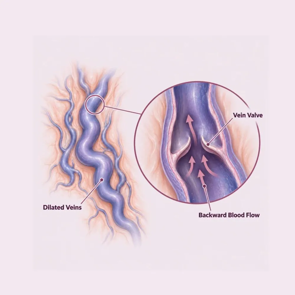
A varicocele is a dilation of the pampiniform plexus, the network of veins that drains blood away from the testicle. Valves within these veins normally keep blood flowing upward toward the heart. When those valves fail, blood pools and the veins distend: the same mechanism that produces varicose veins in the leg.
Around ninety per cent occur on the left side, and that is anatomy rather than coincidence. The left testicular vein drains into the left renal vein at a right angle, a longer and higher-pressure route than the right, which drains directly into the vena cava at a shallow angle. The left side is simply working against more resistance.
The clinical significance is heat. Pooled blood raises the temperature within the scrotum, and sperm production is exquisitely temperature-sensitive: it is the reason the testes sit outside the body in the first place. A varicocele can therefore reduce sperm count, motility and morphology, and in adolescents can impair testicular growth.
The underlying mechanism is valve failure within the testicular veins, and its anatomical basis explains the left-sided predominance.
The valves that keep blood flowing upward stop working, allowing it to reflux and pool. The core mechanism in almost every case.
The left testicular vein enters the left renal vein at a right angle and against higher pressure. This is why ninety per cent of varicoceles are left-sided.
Most varicoceles appear between fifteen and twenty-five, when blood flow to the testes increases sharply during growth.
Chronic straining, heavy lifting and prolonged standing raise venous pressure and can contribute.
Compression of the left renal vein between the aorta and superior mesenteric artery raises pressure downstream into the testicular vein.
Rarely, a mass obstructing venous drainage causes a varicocele: the reason a new, right-sided or non-reducible one is imaged.
Varicocele is a mechanical problem of drainage, not an infection or a lifestyle consequence. Nothing you did caused it, and no medication reverses it.
Most varicoceles cause nothing at all and are found during examination for something else, often an infertility workup.
Common symptoms
Signs that need assessment
An isolated right-sided varicocele, or one that persists when you lie flat, is unusual and warrants abdominal imaging: occasionally it reflects something obstructing the vein higher up.
The diagnosis is clinical, confirmed on ultrasound, and completed with a semen analysis where fertility is a concern.
Varicoceles are examined standing, since they often disappear when supine. Grade one is palpable only on straining, grade two palpable at rest, grade three visible through the skin.
Confirms the diagnosis, measures vein diameter and demonstrates reflux on Valsalva. It also measures testicular volume, which guides treatment in adolescents.
Count, motility and morphology, ideally on two samples. This is what determines whether a symptomless varicocele needs repair.
FSH, LH and testosterone where the semen analysis is abnormal, to assess overall testicular function.
Reserved for an isolated right-sided varicocele, sudden onset, or one that does not reduce on lying flat: to exclude an obstructing mass.
In adolescents, a difference of more than twenty per cent between the two sides is a recognised indication for repair.
A varicocele found incidentally in a man with normal semen parameters and no pain generally needs no treatment at all. The investigations exist to identify the minority in whom it is doing harm.
Treatment is indicated for pain, impaired fertility, or testicular growth failure in adolescents, not for the mere presence of a varicocele.
Appropriate for a symptomless varicocele with normal semen parameters. Periodic semen analysis is all that is needed.
Supportive underwear reduces the dragging ache, particularly for men who stand for long periods.
Anti-inflammatories for intermittent discomfort. Useful for symptom control but does not alter the varicocele.
Avoiding prolonged standing and heavy straining where these clearly aggravate the ache.
The veins are tied off under an operating microscope through a small incision below the groin. The gold standard, with the lowest recurrence and lowest risk of hydrocele or arterial injury.
The testicular veins are clipped high in the abdomen through keyhole ports. Useful for bilateral repair in one sitting.
The traditional approach through a groin incision without a microscope. Effective, but with higher recurrence than the microsurgical technique.
A radiologist passes a catheter through a vein and blocks the testicular vein with coils. No incision, but recurrence is somewhat higher than microsurgery.
Microsurgical repair is the reference standard because the microscope allows the surgeon to identify and spare the testicular artery and the lymphatics, which is what keeps hydrocele and recurrence rates low.
The operation takes around forty-five to ninety minutes depending on whether one or both sides are repaired.
Day-care microsurgery under spinal or general anaesthesia
Fasting from midnight, with routine bloods and anaesthetic review completed beforehand. A single antibiotic dose is given at induction.
Spinal or general anaesthesia. Spinal is common for a single-sided repair; general is usual for bilateral or laparoscopic surgery.
A small incision of two to three centimetres below the groin crease, low enough to avoid dividing the abdominal muscle layers.
The cord is brought out through the incision and supported so its contents can be dissected under magnification.
Under the operating microscope, the surgeon identifies and preserves the testicular artery, the lymphatics and the vas deferens, and isolates the dilated veins individually.
Each dilated vein is tied and divided. Preserving lymphatics is what prevents post-operative hydrocele, and preserving the artery is what protects the testis.
The cord is returned, the layers closed with absorbable sutures, and a scrotal support applied. You go home the same day.
The whole rationale for the microscope is that the testicular artery is roughly a millimetre across and lies among the veins. Without magnification it can be injured, and with it, it very rarely is.
Varicocele surgery is technically demanding at millimetre scale, and the technique determines the outcome. Here is why patients trust us:
Repair under the operating microscope, sparing the testicular artery and lymphatics: the approach with the lowest recurrence and hydrocele rates.
Semen analysis before and after, with a realistic account of expected improvement rather than a promise of one.
From the first consultation through to post-treatment follow-up, with one team throughout.
Our hospital is in HITEC CITY, near IKEA: easily reachable from across Hyderabad.
Physical recovery is quick. The fertility benefit, if that is why you had surgery, takes months to appear.
Home the same day. Mild soreness at the incision, managed with simple painkillers. Scrotal support worn continuously.
Discomfort settles quickly. Walking encouraged; prolonged standing and lifting avoided.
Desk work resumes within a week. The wound is reviewed and the dressing removed.
Light exercise and sexual activity resume. Any residual scrotal swelling settles.
Repeat semen analysis. Improvement in count and motility typically appears at three months and continues to six, because a full sperm production cycle takes about seventy-four days.
Supportive underwear for two to four weeks reduces swelling and discomfort more effectively than anything else.
Straining raises venous pressure at exactly the point that has been repaired. Walking is fine and encouraged.
Testing earlier is not informative, since sperm produced before surgery are still in the system. Three and six months are the meaningful points.
A sperm production cycle takes around seventy-four days. Improvement in parameters appears over months, not weeks.
Visible veins reduce gradually over weeks. They may still be faintly palpable and that does not mean the repair has failed.
A tense, painful scrotum, fever or wound discharge needs review: these suggest haematoma or infection rather than normal healing.
Semen parameters improve in roughly sixty to seventy per cent of men after repair, and pregnancy rates improve correspondingly. It is not a guarantee, and that is discussed honestly before you decide.
Book a consultation if any of the following apply.
Varicocelectomy is day-care surgery. The main variables are the technique used and whether one or both sides are repaired.
| Procedure | Estimated Cost |
|---|---|
| Consultation Includes standing examination | ₹800 – ₹1,500 |
| Scrotal Doppler ultrasound Confirms reflux and measures volumes | ₹2,000 – ₹4,000 |
| Semen analysis Usually two samples | ₹800 – ₹2,000 |
| Microsurgical varicocelectomy: one side Day care, all-inclusive | ₹60,000 – ₹95,000 |
| Microsurgical varicocelectomy: both sides Done in one sitting | ₹85,000 – ₹1,30,000 |
| Laparoscopic varicocelectomy Includes consumables | ₹70,000 – ₹1,10,000 |
| Percutaneous embolisation Dominated by coil and catheter costs | ₹75,000 – ₹1,40,000 |
Microsurgery requires an operating microscope and longer theatre time. Embolisation cost is driven largely by the coils used.
Bilateral repair costs more than a single side but far less than two separate admissions.
Semen analysis and hormone testing add to the assessment cost where infertility is the reason for referral.
A shared ward, a twin-sharing room or a private suite: the largest single swing in most estimates.
We are empanelled with most major insurers, and our team files the pre-authorisation for you.
Every figure above is an indicative range, not a quote. After your consultation you are given a written, fixed-cost estimate, and that is the number you pay, with no additions once treatment begins.
The urologists who perform varicocele surgery at our HITEC CITY hospital.

Consultant Urologist & Andrologist
MBBS, MS, DrNB (Urology)
10 years experience
Consultant Urologist & Andrologist
MBBS, MS, MCh (Urology)
9 years experience
Reviews from patients treated for urological and men’s health conditions at Lux Hospitals, Hyderabad.
Written by Dr. Vipin Reddy B on men’s health and urological care.
Dr. Vipin Reddy B

Dr. Vipin Reddy B

Dr. Vipin Reddy B
Dr. Vipin Reddy B

Dr. Vipin Reddy B

Dr. Vipin Reddy B
Common questions about varicocele, fertility, surgery and what treatment costs at Lux Hospitals.
Still have questions? Call us at 079 6908 4422
Still have questions? Message us on WhatsApp at 079 6908 4444
Doppler assessment, semen analysis and microsurgical varicocele repair with our urologists, with costs shared upfront.
Plot No.116, Lumbini Avenue, HITEC CITY, Near IKEA, Hyderabad – 500032