
Dr. Samhitha Reddy
Consultant Proctologist & Laparoscopic Surgeon
MBBS, MS, FMAS, FISCP, DMAS
- 8 Yrs
- ASCRS Member
Most liver cysts are found by accident, on a scan done for something else entirely, and most need nothing done at all. A simple liver cyst is benign, causes no symptoms, and can safely be left alone for life.
The ones that matter are the large, the symptomatic and the parasitic. Our hepatobiliary team at Lux Hospitals, HITEC CITY distinguishes them on imaging and treats those that need it laparoscopically, while reassuring the majority who genuinely need only observation.
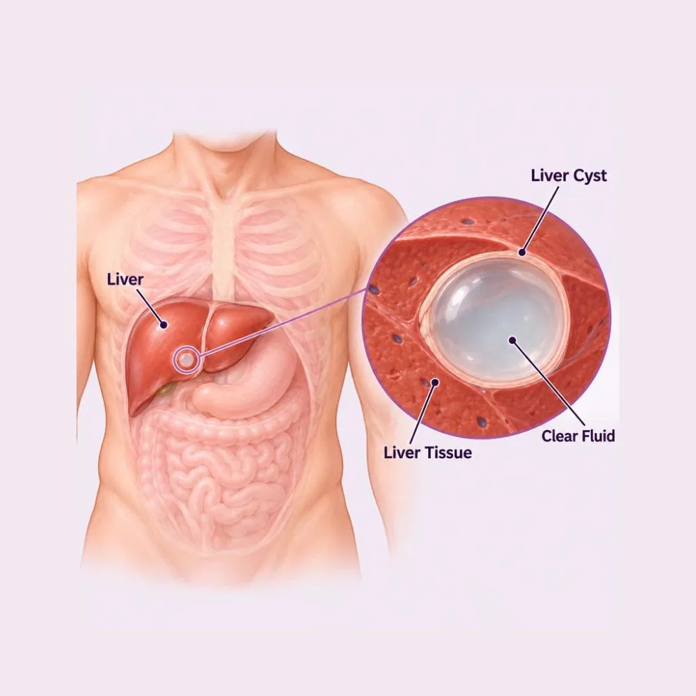
A liver cyst is a fluid-filled sac within the liver tissue. The great majority are simple cysts: thin-walled, containing clear fluid, present from birth and entirely harmless. They are common, they are usually found incidentally, and most people who have one never know.
Several other types exist and behave very differently. A hydatid cyst is caused by a tapeworm parasite and needs specific treatment, handled with particular care because spilling its contents can cause a severe allergic reaction. Polycystic liver disease produces many cysts and is inherited. A cystadenoma is a rare tumour that can become malignant and must be removed completely rather than drained.
Distinguishing these on imaging is the entire clinical task. A simple cyst needs nothing; a cystadenoma needs full excision; a hydatid cyst needs medication before and after surgery. Getting that distinction right is what determines whether treatment is safe.
Different cyst types have entirely different origins, which is why they behave and are treated so differently.
Simple cysts arise from bile ducts that failed to connect properly during development. They are present from birth and grow very slowly.
Echinococcus tapeworm eggs, usually acquired from contact with dogs or contaminated food, form cysts in the liver over years.
An inherited condition producing many cysts, often alongside polycystic kidney disease. It runs strongly in families.
Caroli disease and choledochal cysts involve abnormal dilatation of the bile ducts themselves, which is distinct from a true liver cyst.
A previous liver injury can leave a fluid collection that persists and becomes walled off as a cyst.
A resolving liver abscess can leave a residual cystic cavity that persists on imaging long after the infection has cleared.
A rare benign tumour arising from bile duct epithelium, which carries a real risk of malignant change and must be excised completely.
Simple cysts are commoner in women, and hormonal factors appear to influence their growth.
Nothing you did caused a simple liver cyst. A hydatid cyst, by contrast, comes from a parasite and carries implications for household contacts, which is one reason establishing the type matters.
Most liver cysts cause no symptoms whatsoever. Those that do have usually grown large enough to press on something.
When symptoms occur
Signs of a complication
A simple cyst that has been the same size for years and causes nothing needs no treatment at all. It is worth being told that clearly, because an incidental finding on a scan is easily worried about far more than it deserves.
The task is not detecting the cyst, that is usually already done, but characterising it. Imaging features determine everything that follows.
Usually the test that finds the cyst. A thin-walled, anechoic cyst with no internal features is almost certainly a simple cyst.
Characterises the cyst wall, internal septations, calcification and any solid component: the features that separate simple from complex.
The best test for complex cysts. It shows the internal structure clearly and defines the relationship to the bile ducts before any surgery.
Blood antibody testing for Echinococcus. Essential before touching any cyst with suspicious features, since aspirating a hydatid cyst can be dangerous.
CA 19-9 and CEA where a cystadenoma or malignancy is suspected, alongside liver function tests.
For cysts of uncertain nature, repeat scanning at intervals. Stability over time is itself reassuring evidence.
A hydatid cyst is never aspirated for diagnosis. Spilling its contents can cause anaphylaxis and seed the parasite through the abdomen, which is why serology comes before any needle.
Most liver cysts need no treatment. Where treatment is required, the approach depends entirely on the type of cyst.
The right answer for most simple cysts. Periodic ultrasound confirms stability, and many patients need nothing beyond initial reassurance.
Albendazole for hydatid cysts, given before and after any intervention to sterilise the cyst and reduce the risk of recurrence.
The cyst is drained under imaging guidance and a sclerosant such as alcohol injected to prevent refilling. Less durable than surgery, and only for simple cysts.
Puncture, Aspiration, Injection and Re-aspiration for hydatid cysts, done under strict antibiotic and anti-parasitic cover in selected cases.
For an infected cyst, usually alongside drainage rather than instead of it.
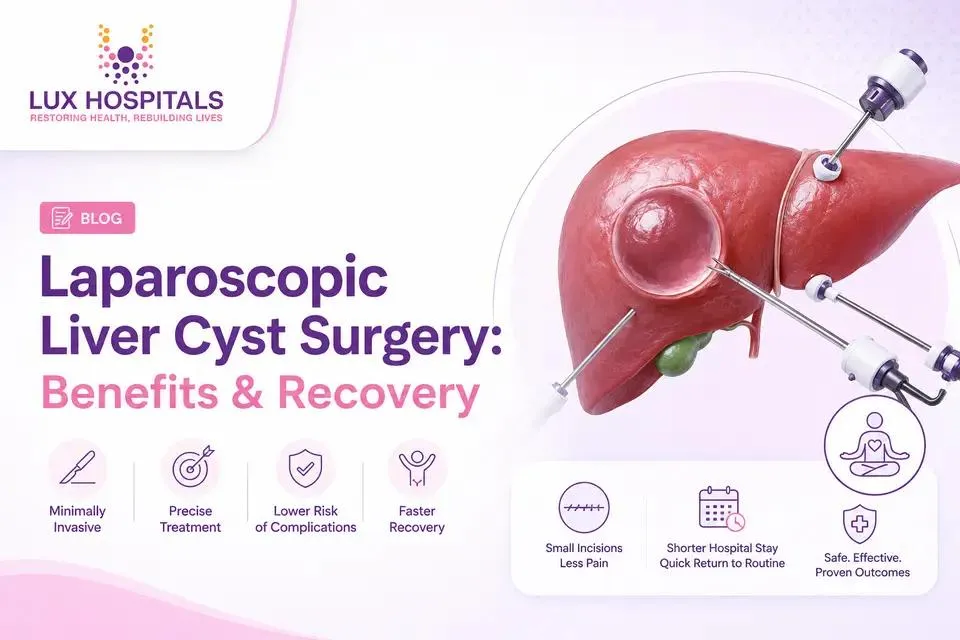
The standard operation for a symptomatic simple cyst. The exposed cyst wall is removed so fluid drains into the abdomen and is absorbed. Excellent results with a short recovery.
Complete removal of the cyst with its wall, required for cystadenoma where leaving any lining behind risks recurrence or malignancy.
Careful removal with the field packed and protected, and scolicidal solution used, so no contents spill. Performed under albendazole cover.
Removal of a segment of liver, for a cystadenoma, suspected malignancy, or extensive disease confined to one part of the liver.
Multiple cysts are deroofed together to relieve pressure symptoms where the liver has become massively enlarged.
Treatment is advised for cysts causing symptoms, cysts that are infected or bleeding, cysts growing rapidly, hydatid cysts, and any cyst with features suggesting a cystadenoma or malignancy. Everything else is watched.
A sixty to ninety minute keyhole operation through three or four small incisions, with a one to two night stay.
Laparoscopic deroofing
Imaging reviewed and hydatid serology confirmed negative. Routine bloods, clotting studies and an anaesthetic review.
General anaesthetic. The abdomen is inflated with carbon dioxide and three or four small ports are placed.
The liver is inspected and the cyst located. Intraoperative ultrasound is used where the cyst is deep or its relationship to vessels is unclear.
The cyst is punctured and drained, and the fluid inspected. Clear fluid supports a simple cyst; bile-stained or turbid fluid changes the plan.
The exposed portion of the cyst wall is excised widely, leaving the cavity open to the abdomen so any further fluid is simply absorbed.
The lining is inspected for solid areas or papillary projections that would suggest a cystadenoma. Any suspicious lining is sent for frozen section.
The wall is sent for histopathology, a drain is occasionally left, and you are home after one to two nights.
Deroofing rather than simple drainage is what makes this durable. A cyst that is only aspirated refills within months, because the secreting lining is still there.
The most valuable thing here is often being told, accurately, that nothing needs doing. Here is why patients trust us:
CT, MRI and hydatid serology, so a simple cyst is confidently distinguished from one needing surgery.
Led by Dr. Tagore Mohan Grandhi, with extensive experience in laparoscopic liver surgery including hydatid disease.
From the first consultation through to post-treatment follow-up, with one team throughout.
Our hospital is in HITEC CITY, near IKEA: easily reachable from across Hyderabad.
Recovery after laparoscopic deroofing takes one to two weeks. Formal liver resection and hydatid surgery take considerably longer.
Overnight in the ward. Sips of fluid the same evening, eating and walking the next morning. Some shoulder-tip discomfort from the gas.
Home once eating, mobile and comfortable on oral painkillers. Port site soreness settles quickly.
Light activity and desk work resume. Driving from around ten days. Dressings removed and wounds checked.
Normal activity including the gym resumes progressively. Histopathology on the cyst wall is discussed at follow-up.
Follow-up ultrasound confirms the cavity has collapsed and there is no recurrence. Hydatid patients continue albendazole and longer surveillance.
Nothing above five kilos for the first month, so the port sites heal without strain.
For hydatid cysts the course continues for months after surgery. Stopping early is the main cause of recurrence.
No special diet is needed after liver cyst surgery. A balanced diet supports healing.
Walking from day one, with a graded return to normal exercise over four to six weeks.
Fever, worsening abdominal pain, jaundice, wound discharge, or itching and rash suggesting a hydatid reaction.
Ultrasound at intervals confirms no recurrence. Hydatid disease needs surveillance for several years.
Recurrence after proper deroofing is uncommon. It is more likely after simple aspiration, because the cyst lining is left behind and continues to secrete.
Treatment is advised if any of the following apply.
Many patients need only a consultation and a scan. These are the ranges for those who need treatment.
| Procedure | Estimated Cost |
|---|---|
| Consultation & assessment Per visit | ₹1,000 – ₹2,000 |
| Ultrasound & CT scan Characterising the cyst | ₹6,000 – ₹15,000 |
| MRI with MRCP For complex cysts | ₹12,000 – ₹22,000 |
| Aspiration with sclerotherapy Day care or one night’s stay | ₹35,000 – ₹65,000 |
| Laparoscopic deroofing One to two nights’ stay | ₹1,40,000 – ₹2,50,000 |
| Hydatid cyst surgery Three to five nights, with albendazole cover | ₹2,00,000 – ₹3,50,000 |
| Liver resection for cystadenoma Five to seven nights’ stay | ₹3,00,000 – ₹5,50,000 |
A simple cyst deroofed laparoscopically and a cystadenoma requiring formal liver resection are entirely different operations.
A cyst deep within the liver or close to major vessels is considerably more demanding than a superficial one.
Hydatid surgery requires albendazole before and after, scolicidal solutions, and meticulous field protection: all of which add to the cost.
A shared ward, a twin-sharing room or a private suite: the largest single swing in most estimates.
We are empanelled with most major insurers, and our team files the pre-authorisation for you.
Every figure above is an indicative range, not a quote. After your consultation you are given a written, fixed-cost estimate, and that is the number you pay, with no additions once treatment begins.
The gastrointestinal and hepatobiliary surgeon who treats liver cysts at our HITEC CITY hospital.

Senior Consultant: Bariatric & Gastrointestinal Surgery
MBBS, MS, FRCS (UK), DLS (France)
28 years experience
Reviews from patients treated for liver and gastrointestinal conditions at Lux Hospitals, Hyderabad.
Written by Dr. Tagore Mohan Grandhi on liver, digestive and gastrointestinal surgical care.

Dr. Tagore Mohan Grandhi

Dr. Tagore Mohan Grandhi
Dr. Tagore Mohan Grandhi
Dr. Tagore Mohan Grandhi
Dr. Tagore Mohan Grandhi
Dr. Tagore Mohan Grandhi
Common questions about liver cysts, their treatment, and what surgery costs at Lux Hospitals.
Still have questions? Call us at 079 6908 4422
Still have questions? Message us on WhatsApp at 079 6908 4444
Accurate characterisation of your liver cyst and treatment only where it is genuinely needed, with costs shared upfront.
Plot No.116, Lumbini Avenue, HITEC CITY, Near IKEA, Hyderabad – 500032