Dr. Samhitha Reddy
Consultant Proctologist & Laparoscopic Surgeon
MBBS, MS, FMAS, FISCP, DMAS
- 8 Yrs
- ASCRS Member
Kidney stone pain is among the most severe pain in medicine: patients routinely describe it as worse than childbirth. It comes in waves, radiates from the loin to the groin, and no position relieves it.
Most stones pass on their own with the right support. For those that do not, our urologists at Lux Hospitals, HITEC CITY use laser lithotripsy, RIRS and keyhole PCNL: techniques that clear stones without a single external incision.
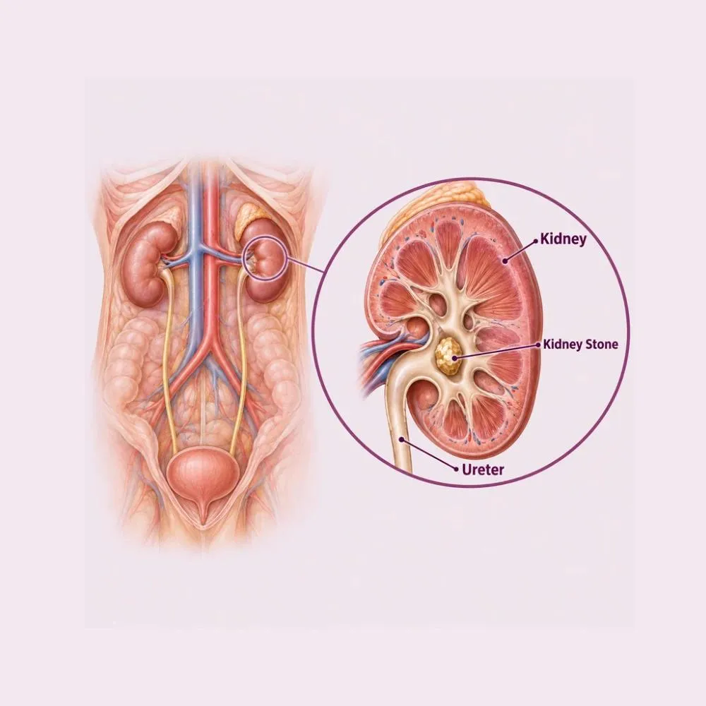
Kidney stones are hard deposits of minerals and salts that crystallise inside the kidney when urine becomes too concentrated. They can sit silently in the kidney for years and cause nothing at all: the trouble begins when one moves into the ureter, the narrow tube draining the kidney, and obstructs it.
The pain of renal colic comes from that obstruction rather than from the stone itself. Urine backs up, the kidney swells, and the ureter goes into spasm trying to push the stone along. It comes in waves, is often accompanied by vomiting, and characteristically nothing you do relieves it.
Stones vary in composition, and that matters for preventing the next one. Calcium oxalate stones account for the great majority. Uric acid stones can sometimes be dissolved with medication alone. Struvite stones form in the presence of infection and can grow very large. Cystine stones are rare and inherited. This is why every stone retrieved is sent for analysis.
Stones form when urine becomes supersaturated with stone-forming minerals. Dehydration is the single largest contributor, particularly in Indian summers.
The commonest and most correctable cause. Concentrated urine allows crystals to form and grow, and hot climates make this considerably worse.
Salt increases calcium excretion into the urine, and dietary salt reduction is one of the most effective preventive measures.
Spinach, nuts, chocolate, tea and beetroot are high in oxalate, which binds calcium in urine to form the commonest stone type.
High intake raises urinary calcium and uric acid while lowering citrate, the body’s natural stone inhibitor.
Associated with more acidic urine and higher excretion of stone-forming substances, and it independently raises the risk.
Stones run in families, reflecting both inherited metabolic tendencies and shared diet and climate.
Hyperparathyroidism, gout, recurrent urinary infections, inflammatory bowel disease and renal tubular acidosis all promote stone formation.
Some diuretics, calcium-based antacids, topiramate and excessive vitamin C supplementation all increase risk.
Someone who has formed one stone has roughly a fifty per cent chance of forming another within five to ten years. That is exactly why prevention matters as much as treating the current stone.
A stone sitting in the kidney often causes nothing. A stone moving down the ureter causes the most distinctive pain in urology.
Typical symptoms
Emergency features: seek care now
Fever with renal colic is a urological emergency. An obstructed kidney with infection above it can cause sepsis within hours, and it needs urgent drainage rather than antibiotics alone.
A non-contrast CT is the definitive test. It shows stone size, position and density: all of which determine which treatment is right.
The gold standard, detecting virtually every stone regardless of composition, with precise size and position. It also measures density, which predicts how the stone will respond to shock waves.
First-line in pregnancy and in children, avoiding radiation. It shows kidney swelling and larger stones, though small ureteric stones are often missed.
Dipstick for blood, and culture to detect infection, which changes an elective problem into an urgent one.
Kidney function, calcium, uric acid and inflammatory markers, assessing both the kidney and any metabolic cause.
Useful for following a known radio-opaque stone over time with much less radiation than repeated CT.
Every retrieved stone is analysed, and recurrent stone formers get a 24-hour urine study to identify the underlying cause.
Stone size predicts what will happen next more than anything else. Stones under five millimetres pass spontaneously in most people; those above ten millimetres rarely do and usually need intervention.
Small stones are given the chance to pass with support. Larger ones are treated with techniques that leave no external incision at all.
For stones under five millimetres, high fluid intake and pain relief. Most pass spontaneously within four weeks.
Alpha blockers such as tamsulosin relax the ureter and improve the chance a stone in the lower ureter will pass.
Anti-inflammatory medication is more effective than opioids for renal colic, since it reduces the ureteric spasm directly.
Uric acid stones can be dissolved by alkalinising the urine with potassium citrate: the one stone type treatable with medication alone.
Focused shock waves from outside the body break the stone into fragments that pass in the urine. No anaesthetic incision, though fragments still have to pass.
A fine scope is passed up the natural urinary passage and a laser fragments the stone, with pieces retrieved directly. No incision, and often a day-care procedure.
Retrograde Intra-Renal Surgery: a flexible scope reaches into the kidney itself through the natural passage, treating stones up to around two centimetres.
Percutaneous nephrolithotomy: a keyhole tract into the kidney through the back for large or staghorn stones. The most effective option for bulky stone burden.
The same principle through a smaller tract, with less bleeding and a shorter stay for moderate-sized stones.
For an infected obstructed kidney, urgent drainage by stent or a tube through the back. Definitive stone treatment follows once the infection has settled.
A ureteric stent is often placed after treatment to keep the ureter open while swelling settles. It causes some urinary urgency and discomfort, which is expected and resolves as soon as it is removed.
A thirty to sixty minute procedure performed entirely through the natural urinary passage. No incision, no external wound, and usually a day case.
Ureteroscopy with laser lithotripsy
CT reviewed for stone size and position, urine culture confirmed clear of infection, and routine bloods. Operating through infected urine risks sepsis.
General or spinal anaesthesia. The procedure is not painful afterwards, though the stent can cause urgency.
A fine ureteroscope is passed through the urethra and bladder and up the ureter, under direct vision and X-ray guidance.
The stone is identified directly. A flexible scope is used where it sits within the kidney rather than the ureter.
A holmium laser fibre is passed down the scope and fragments the stone into dust or small pieces. The laser works on every stone composition.
Pieces are removed with a basket rather than left to pass, and one is always sent for composition analysis.
A stent is usually left for one to two weeks, and you go home the same day or the next morning.
Sending a fragment for analysis is not a formality. Knowing whether the stone was calcium oxalate, uric acid or struvite is what makes it possible to prevent the next one specifically rather than generically.
Stone treatment is now almost entirely incision-free, and preventing the next one matters as much as clearing this one. Here is why patients trust us:
A dedicated urology team performing laser lithotripsy, RIRS and PCNL routinely, with the full range of techniques available.
Stone analysis and metabolic assessment for recurrent formers, so the next stone is prevented rather than just awaited.
From the first consultation through to post-treatment follow-up, with one team throughout.
Our hospital is in HITEC CITY, near IKEA: easily reachable from across Hyderabad.
Recovery from endoscopic stone surgery is quick. Preventing the next stone is the part that takes ongoing effort, and it works.
Home the same day or the next morning after ureteroscopy. Some burning on passing urine and blood in the urine, both expected.
Normal activity resumes. Stent symptoms (urgency, frequency and mild flank discomfort on passing urine) are common and settle.
Stent removed in the clinic, a quick procedure needing no anaesthetic. Symptoms resolve immediately afterwards.
Full recovery. After PCNL, the small back wound heals and normal activity resumes over this period.
Follow-up imaging confirms the kidney is clear, and stone analysis and metabolic results guide a prevention plan.
The single most effective prevention there is. Aim for pale urine throughout the day, and more in summer or if you work outdoors.
Salt drives calcium into the urine. Cutting it is often more effective than cutting calcium, which should not be restricted.
A common and counterproductive mistake. Normal dietary calcium binds oxalate in the gut and actually reduces stone risk.
Lemon juice and citrus inhibit stone formation naturally. Potassium citrate is prescribed where a metabolic study indicates it.
Fever, uncontrolled pain, inability to pass urine, or heavy bleeding. Fever with a stent or a stone always needs same-day assessment.
A stent left in beyond its intended period encrusts with stone and becomes difficult to remove. Never miss that appointment.
Around half of stone formers make another within five to ten years, but that figure falls substantially with adequate fluid intake, salt reduction and treatment of any metabolic abnormality found.
Come in promptly for the first three, and go to the emergency department for the rest.
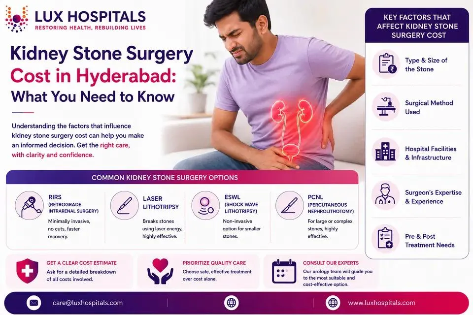
Cost depends on stone size, position and the technique used. These are the ranges our patients fall into.
| Procedure | Estimated Cost |
|---|---|
| Consultation & assessment Per visit | ₹800 – ₹1,500 |
| CT KUB scan The definitive diagnostic test | ₹4,000 – ₹9,000 |
| Medical management Pain relief and expulsive therapy for small stones | ₹2,000 – ₹6,000 |
| Shock wave lithotripsy (ESWL) Per session; may need repeating | ₹25,000 – ₹55,000 |
| Ureteroscopy with laser (URSL) Day care or one night; stent included | ₹55,000 – ₹1,10,000 |
| RIRS One night’s stay; flexible scope and laser | ₹85,000 – ₹1,70,000 |
| PCNL Two to four nights; for large stones | ₹1,20,000 – ₹2,40,000 |
A small ureteric stone treated by ureteroscopy and a staghorn stone needing PCNL are entirely different procedures.
Stones in both kidneys, or several in one, may need staged procedures a few weeks apart.
An infected obstructed kidney needs urgent drainage first, then definitive treatment later: two admissions rather than one.
A shared ward, a twin-sharing room or a private suite: the largest single swing in most estimates.
We are empanelled with most major insurers, and our team files the pre-authorisation for you.
Every figure above is an indicative range, not a quote. After your consultation you are given a written, fixed-cost estimate, and that is the number you pay, with no additions once treatment begins.
What patients treated here can expect:
Treatment through the natural urinary passage, leaving no external wound.
Day-care options with a return to normal activity within days.
Stones fragmented and retrieved rather than left to pass on their own.
The urologists who treat kidney stones at our HITEC CITY hospital.

Consultant Urologist & Andrologist
MBBS, MS, DrNB (Urology)
10 years experience

Consultant Urologist & Andrologist
MBBS, MS, MCh (Urology)
9 years experience
Reviews from patients treated for kidney stones and urological conditions at Lux Hospitals, Hyderabad.
Written by Dr. Vipin Reddy B on kidney stones, urinary health and urological care.
Dr. Vipin Reddy B
Dr. Vipin Reddy B
Dr. Vipin Reddy B
Dr. Vipin Reddy B
Dr. Vipin Reddy B

Dr. Vipin Reddy B
Common questions about kidney stones, laser treatment, and what surgery costs at Lux Hospitals.
Still have questions? Call us at 079 6908 4422
Still have questions? Message us on WhatsApp at 079 6908 4444
Incision-free laser stone treatment with a prevention plan afterwards, and a fixed-cost estimate before treatment begins.
Plot No.116, Lumbini Avenue, HITEC CITY, Near IKEA, Hyderabad – 500032