
Dr. Samhitha Reddy
Consultant Proctologist & Laparoscopic Surgeon
MBBS, MS, FMAS, FISCP, DMAS
- 8 Yrs
- ASCRS Member
A pancreatic pseudocyst is a collection of pancreatic fluid that forms after an attack of pancreatitis or an injury to the pancreas. Many resolve on their own over weeks, and the temptation to drain them early is one worth resisting.
Timing is the whole art here. Our pancreatic team at Lux Hospitals, HITEC CITY monitors those that are likely to settle and drains the ones that are not: using endoscopic ultrasound-guided drainage through the stomach wall, which avoids an operation entirely in most cases.
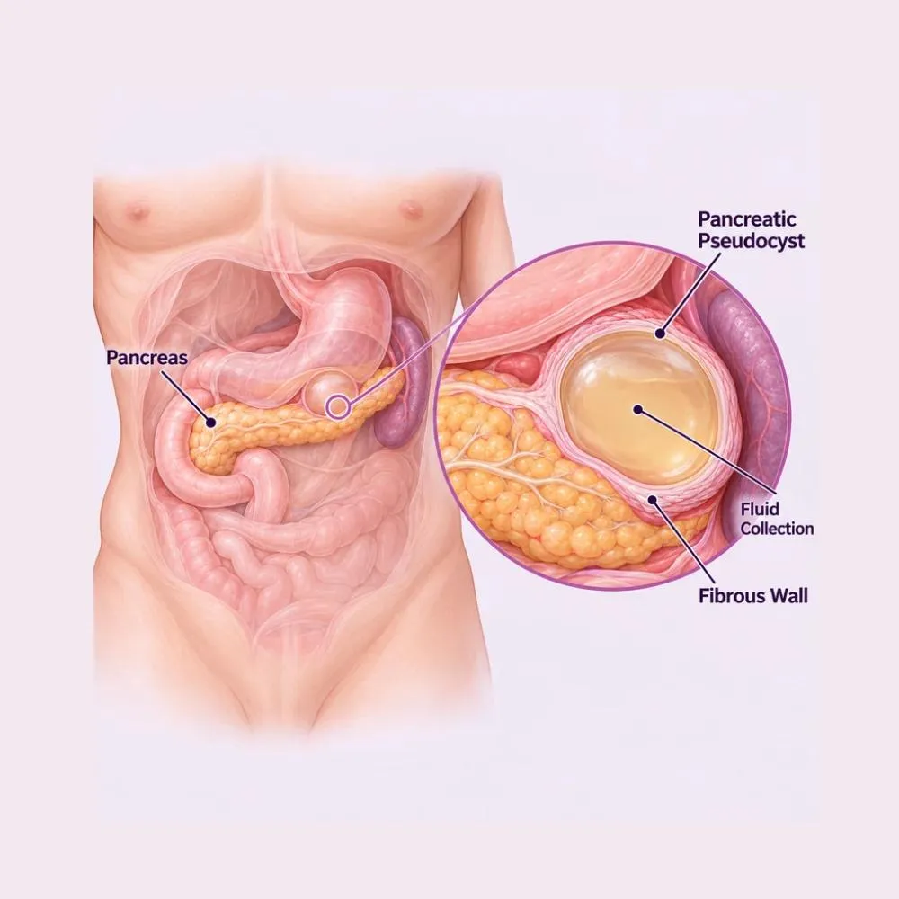
A pancreatic pseudocyst is a collection of pancreatic fluid enclosed by a wall of fibrous and inflammatory tissue. It is called a pseudocyst (a false cyst) because that wall is scar tissue rather than the epithelial lining a true cyst has. That distinction matters, because it determines how the collection behaves and how it can be treated.
Pseudocysts form when the pancreatic duct is disrupted, usually by an attack of pancreatitis or by injury, and enzyme-rich fluid leaks out and becomes walled off. The wall takes around four weeks to mature, which is the single most important fact governing treatment: a collection that has not yet developed a firm wall cannot be drained safely.
Their natural history is favourable. A significant proportion of pseudocysts resolve spontaneously over weeks to months as the fluid is reabsorbed, particularly those under six centimetres. This is why observation is often the right initial approach rather than a failure to act.
Every pseudocyst begins with disruption of the pancreatic duct, allowing enzyme-rich fluid to escape and become walled off.
The commonest cause. Inflammation disrupts the duct and fluid collects, typically declaring itself around four weeks after the attack.
Duct strictures and stones cause raised pressure upstream, leading to leakage and pseudocyst formation, often recurrently.
A blunt injury to the upper abdomen, a handlebar or seatbelt injury, can transect the pancreatic duct and cause a pseudocyst weeks later.
A stone, stricture or tumour blocking the duct causes back-pressure that eventually forces fluid out into the surrounding tissue.
The commonest underlying context in adults, and the group most likely to develop recurrent pseudocysts if drinking continues.
A frequent cause of the initial attack, and one where removing the gallbladder prevents both further pancreatitis and further pseudocysts.
Damage to the pancreatic duct during surgery on the stomach, spleen or pancreas can result in a collection afterwards.
Uncommonly, an endoscopic procedure on the pancreatic duct can cause a leak leading to a pseudocyst.
Treating the pseudocyst without addressing the underlying duct problem invites recurrence. Where a stricture or stone is present, dealing with it is part of the treatment rather than a separate matter.
Small pseudocysts often cause nothing at all. Larger ones cause symptoms by pressing on the stomach and surrounding structures.
Common symptoms
Signs of a complication
Sudden severe pain, fever, or bleeding in someone known to have a pseudocyst needs emergency assessment. Rupture, infection and haemorrhage are the three complications that turn a watchable problem into an urgent one.
Imaging confirms the collection and its maturity. The critical task is distinguishing a pseudocyst from a cystic neoplasm, which needs entirely different treatment.
Usually the first test. It detects the collection, measures it, and is convenient for monitoring change over time.
Defines size, position, wall maturity and the relationship to the stomach: all of which determine whether and how it can be drained.
Shows the pancreatic duct and whether it communicates with the collection, and distinguishes fluid from solid debris better than CT.
The definitive test. High amylase with low CEA in the fluid confirms a pseudocyst; high CEA indicates a mucinous neoplasm instead.
Amylase, lipase, full blood count and inflammatory markers, plus liver function where bile duct compression is suspected.
Repeat scans over weeks track whether the collection is resolving, stable or enlarging, which is what guides the decision to intervene.
Mistaking a mucinous cystic neoplasm for a pseudocyst and draining it is a serious error: it would leave a premalignant lesion in place. Cyst fluid analysis is what prevents that.
Many pseudocysts need no treatment at all. Where drainage is required, endoscopic approaches have largely replaced surgery.
The right approach for asymptomatic pseudocysts, particularly those under six centimetres. A significant proportion resolve on their own over weeks to months.
Adequate nutrition, sometimes by nasojejunal tube, to allow the pancreas to rest while the collection settles.
Adequate analgesia while awaiting resolution, avoiding NSAIDs where there is any bleeding risk.
Stopping alcohol, removing the gallbladder after gallstone pancreatitis, and correcting raised triglycerides.
The treatment of choice. A stent is placed between the stomach and the pseudocyst under endoscopic ultrasound guidance, draining it internally with no incision at all.
Wide stents that hold the connection open and allow debris to pass, and permit an endoscope to be passed in later if necrosis needs clearing.
A stent placed through the pancreatic duct at ERCP, suitable where the collection communicates directly with the duct.
An external drain placed under CT or ultrasound guidance. Effective, but it carries a risk of creating a persistent external pancreatic fistula.
A surgical join between the pseudocyst and the stomach, laparoscopic or open. Reserved for collections not accessible endoscopically or where endoscopic drainage has failed.
Removal of the tail of the pancreas with the pseudocyst, where there is duct disruption in the tail or diagnostic uncertainty.
Drainage is indicated for pseudocysts that are symptomatic, larger than six centimetres, enlarging, infected, or compressing the stomach or bile duct. Size alone in an asymptomatic patient is not automatically a reason to intervene.
A forty-five to sixty minute endoscopic procedure done through the mouth. No incisions, no external drain, and usually a one to two night stay.
Endoscopic ultrasound-guided cystgastrostomy
Imaging confirms the wall has matured: at least four weeks from the causative attack. Draining an immature collection risks leakage into the abdomen.
General anaesthetic or deep sedation with the airway protected. The endoscope is passed into the stomach.
Endoscopic ultrasound identifies the pseudocyst through the stomach wall and, crucially, maps the intervening blood vessels so they can be avoided.
A needle is passed from the stomach into the collection under real-time ultrasound, and fluid aspirated for analysis to confirm the diagnosis.
A guidewire is coiled inside the cavity and a stent, often a lumen-apposing metal stent, deployed to hold the connection open.
Fluid is seen draining into the stomach. Where solid debris is present, the endoscope can be passed through the stent to clear it directly.
One to two nights in hospital, then home. Follow-up imaging confirms resolution, and the stent is removed endoscopically after four to eight weeks.
Draining the collection internally into the stomach is what avoids an external fistula. A percutaneous drain works, but it can leave a tract that continues to leak pancreatic fluid for months.
The judgement here is about timing and about knowing which collections will settle on their own. Here is why patients trust us:
Endoscopic ultrasound-guided stent drainage as the default, avoiding external drains and the fistulas they can leave behind.
Led by Dr. Tagore Mohan Grandhi, with extensive experience in pancreatic collections and their surgical management.
From the first consultation through to post-treatment follow-up, with one team throughout.
Our hospital is in HITEC CITY, near IKEA: easily reachable from across Hyderabad.
Recovery after endoscopic drainage is quick. The collection itself takes weeks to resolve fully, and the stent stays in during that period.
Where the collection is being watched, serial imaging tracks whether it is resolving. Many settle without any intervention.
One to two nights in hospital. Symptoms of pressure and fullness typically improve immediately once the collection is decompressed.
Home on a low-fat diet. Appetite improves and pain settles. Repeat imaging confirms the collection is shrinking.
Follow-up imaging confirms resolution, and the stent is removed endoscopically once the cavity has collapsed.
The underlying cause is addressed (gallbladder removal, alcohol cessation, or treatment of a duct stricture) to prevent recurrence.
Where alcohol caused the pancreatitis, continued drinking leads to further attacks and further pseudocysts.
Small, low-fat meals reduce pancreatic stimulation while the collection resolves. Normal diet resumes gradually afterwards.
Gallbladder removal after gallstone pancreatitis, or clearing a duct stricture. Without this, recurrence is likely.
The stent must be removed once the collection has resolved. Leaving it indefinitely risks bleeding and migration.
Fever, sudden severe abdominal pain, vomiting blood, black stools, or a return of the fullness and pain that had settled.
Repeat scans confirm resolution and detect recurrence early, when it is still straightforward to manage.
Recurrence is uncommon where the underlying cause has been addressed, and common where it has not. The gallbladder, the alcohol or the duct stricture is what determines which group you are in.
Book a consultation for the first three, and seek emergency care for the rest.
Many patients need only monitoring. These are the ranges for those requiring intervention.
| Procedure | Estimated Cost |
|---|---|
| Consultation & assessment Per visit | ₹1,000 – ₹2,000 |
| CT and MRI imaging Characterising the collection | ₹15,000 – ₹35,000 |
| Endoscopic ultrasound with fluid analysis Confirms diagnosis and excludes neoplasm | ₹25,000 – ₹50,000 |
| Percutaneous drainage Three to seven nights’ stay | ₹60,000 – ₹1,20,000 |
| EUS-guided stent drainage One to two nights; stent cost included | ₹1,50,000 – ₹3,00,000 |
| Laparoscopic cystgastrostomy Five to seven nights’ stay | ₹2,50,000 – ₹4,50,000 |
| Distal pancreatectomy Seven to ten nights’ stay | ₹3,50,000 – ₹6,00,000 |
A significant proportion of pseudocysts resolve on their own with observation, at the cost of a few scans and consultations.
Lumen-apposing metal stents cost considerably more than plastic ones, but drain debris far better and allow later necrosectomy.
Walled-off necrosis with solid debris usually needs several endoscopic sessions rather than a single drainage.
A shared ward, a twin-sharing room or a private suite: the largest single swing in most estimates.
We are empanelled with most major insurers, and our team files the pre-authorisation for you.
Every figure above is an indicative range, not a quote. After your consultation you are given a written, fixed-cost estimate, and that is the number you pay, with no additions once treatment begins.
The gastrointestinal and pancreatic surgeon who treats pancreatic pseudocysts at our HITEC CITY hospital.

Senior Consultant: Bariatric & Gastrointestinal Surgery
MBBS, MS, FRCS (UK), DLS (France)
28 years experience
Reviews from patients treated for pancreatic and gastrointestinal conditions at Lux Hospitals, Hyderabad.
Written by Dr. Tagore Mohan Grandhi on pancreatic and digestive health.

Dr. Tagore Mohan Grandhi

Dr. Tagore Mohan Grandhi
Dr. Tagore Mohan Grandhi
Dr. Tagore Mohan Grandhi
Dr. Tagore Mohan Grandhi
Dr. Tagore Mohan Grandhi
Common questions about pancreatic pseudocysts, their drainage, and what treatment costs at Lux Hospitals.
Still have questions? Call us at 079 6908 4422
Still have questions? Message us on WhatsApp at 079 6908 4444
Accurate assessment and correctly timed drainage of pancreatic collections, with costs shared before treatment begins.
Plot No.116, Lumbini Avenue, HITEC CITY, Near IKEA, Hyderabad – 500032