
Dr. Samhitha Reddy
Consultant Proctologist & Laparoscopic Surgeon
MBBS, MS, FMAS, FISCP, DMAS
- 8 Yrs
- ASCRS Member
A liver abscess is a collection of pus inside the liver. It is a serious infection that becomes dangerous if left untreated, and it is one of the conditions where the difference between treating early and treating late is measured in outcomes rather than in convenience.
Most abscesses are cured with antibiotics and image-guided drainage: no open surgery at all. Our gastrointestinal team at Lux Hospitals, HITEC CITY diagnoses with ultrasound and CT, drains through a needle or catheter, and identifies the underlying source that caused it.
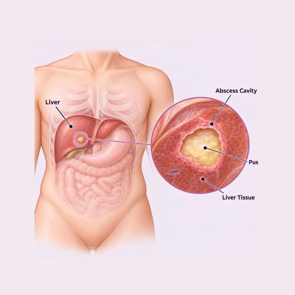
A liver abscess, also called a hepatic abscess, is a walled-off collection of pus within the liver tissue, formed when infection reaches the liver and the body contains it. Untreated it can enlarge, rupture, or seed infection into the bloodstream.
There are two main types, and distinguishing them changes the treatment entirely. An amoebic liver abscess is caused by the parasite Entamoeba histolytica, is usually a single large abscess in the right lobe, and responds dramatically to anti-parasitic medication, often without any drainage at all. A pyogenic abscess is bacterial, is more often multiple, and usually needs both antibiotics and drainage.
In India, amoebic abscesses are considerably more common than in Western practice, which shapes how these are approached here. A large right-lobe abscess in a patient with a history of diarrhoea is amoebic until proved otherwise, and the treatment response is usually the confirmation.
Infection reaches the liver by one of several routes, and identifying which one matters, because the source usually needs treating too.
Entamoeba histolytica travels from the intestine to the liver via the portal vein. The commonest cause in India, and it responds dramatically to medication.
The commonest route for bacterial abscesses. Stones or a stricture obstruct the bile ducts, and infection ascends into the liver.
Appendicitis, diverticulitis or an intestinal perforation seeds bacteria into the portal circulation, which drains directly to the liver.
Bacteria from a distant infection reach the liver through the hepatic artery, particularly in patients with reduced immunity.
A major risk factor, and associated with a particularly aggressive form of Klebsiella liver abscess that can spread to the eyes and brain.
Injury to the liver, or a previous liver or biliary operation, can leave a collection that becomes infected.
Chemotherapy, long-term steroids, HIV or chronic liver disease all raise the risk and can blunt the usual symptoms.
A parasitic hydatid cyst that becomes secondarily infected, which needs quite different handling to avoid spilling its contents.
Finding the source is part of the treatment. An abscess drained without addressing an obstructed bile duct or an inflamed appendix will simply re-form.
The classic combination is fever with right upper abdominal pain, developing over days to weeks rather than suddenly.
Common symptoms
Signs of a serious complication
Fever with right upper abdominal pain for more than a few days needs an ultrasound, not a further course of oral antibiotics. Liver abscesses are frequently treated as unexplained fever for weeks before the scan is done.
Ultrasound usually makes the diagnosis within minutes. The rest of the workup establishes the type, the source and the severity.
The first-line test. It detects the abscess, measures it, shows how many there are, and guides aspiration at the same visit.
More precise than ultrasound for size, number and position, and it identifies the underlying source: an inflamed appendix, diverticulitis or biliary obstruction.
Raised white cells and inflammatory markers, liver function tests, blood sugar to detect undiagnosed diabetes, and blood cultures.
Antibody testing for Entamoeba histolytica. A positive result strongly supports an amoebic abscess and often avoids any drainage at all.
Pus is sent for culture and sensitivity. Anchovy-sauce-coloured fluid without organisms suggests amoebic; frank pus with bacteria suggests pyogenic.
Often shows a raised right diaphragm or a reactive pleural effusion, and helps assess whether the abscess has affected the chest.
A blood sugar check is done on every patient with a liver abscess. Undiagnosed diabetes turns up often enough here that it is a routine part of the workup rather than an afterthought.
Most liver abscesses are cured without any operation. Antibiotics or anti-parasitic drugs, combined with image-guided drainage where needed, resolve the great majority.
Broad-spectrum antibiotics started immediately, then narrowed once culture results are available. Typically two to three weeks intravenously followed by oral therapy.
Metronidazole for amoebic abscess, followed by a luminal agent to clear the intestine. The response is often dramatic, with fever settling within days.
Intravenous fluids, nutritional support, pain relief and correction of anaemia: important in patients who have been unwell for weeks.
Tight blood sugar management, since poorly controlled diabetes both causes these abscesses and impedes their resolution.
A needle is passed into the abscess under ultrasound guidance and the pus withdrawn. Done under local anaesthetic, and often sufficient on its own for smaller abscesses.
A small drain is left in place for continuous drainage over several days. Preferred for larger abscesses that would otherwise re-accumulate.
Where an obstructed bile duct is the cause, clearing it endoscopically treats the source rather than just the collection.
Keyhole surgery for abscesses in positions that cannot be reached percutaneously, or where percutaneous drainage has failed.
Reserved for a ruptured abscess, multiple large abscesses, or where the underlying source needs surgical treatment at the same time.
Surgery is now the exception rather than the rule. The combination of appropriate drugs and image-guided drainage resolves the large majority, and open drainage is reserved for rupture or failure of everything else.
A twenty to thirty minute bedside or radiology-suite procedure under local anaesthetic and ultrasound guidance. No general anaesthetic and no incision.
Percutaneous catheter drainage
Clotting studies and platelet count, since the liver is a vascular organ. Antibiotics are already running before the needle goes in.
You lie on your back or slightly turned. Ultrasound identifies the abscess and plans a route that avoids lung, bowel and major vessels.
The skin and the tissue down to the liver capsule are numbed. The liver itself has no pain fibres, so it is only the surface that needs anaesthetising.
A needle is advanced into the abscess under real-time ultrasound and pus is aspirated. Its appearance is often diagnostic in itself, and it goes for culture.
For larger abscesses a fine catheter is threaded over a guidewire into the cavity and secured to the skin, so it drains continuously.
Imaging confirms the drain sits within the cavity and that the abscess has collapsed around it.
Drain output is recorded daily. Once output falls below around ten millilitres a day and a scan shows resolution, the drain is removed.
The drain typically stays in for five to fourteen days. Removing it too early is the commonest reason an abscess re-accumulates, so the timing is guided by output and imaging rather than by how well you feel.
A liver abscess needs prompt imaging, the right drug for the right organism, and drainage by someone who does it often. Here is why patients trust us:
Same-day ultrasound and CT, with drainage arranged immediately rather than after a further trial of oral antibiotics.
Led by Dr. Tagore Mohan Grandhi, with extensive experience in hepatobiliary conditions and image-guided drainage.
From the first consultation through to post-treatment follow-up, with one team throughout.
Our hospital is in HITEC CITY, near IKEA: easily reachable from across Hyderabad.
Fever usually settles within days of appropriate treatment. The abscess cavity itself takes weeks to months to disappear entirely on imaging.
Intravenous antibiotics or anti-parasitic medication started, with drainage where indicated. Fever typically begins settling within 48 to 72 hours.
In hospital on intravenous treatment with the drain in place. Appetite and energy improve steadily. Drain output is monitored daily.
Drain removed once output is minimal and imaging shows resolution. Home on oral antibiotics to complete the course.
Oral antibiotics completed. Energy returns and lost weight begins to be regained. Repeat ultrasound to confirm the cavity is shrinking.
The cavity resolves completely on imaging. Full recovery for most patients, with the underlying source addressed by this point.
Four to six weeks in total is common. Stopping early because you feel better is the main reason abscesses recur.
A high-protein diet and nutritional support. Most patients have lost weight and muscle over weeks of illness.
If diabetes was found or is known, tight control is essential: it both caused the abscess and slows its resolution.
Fatigue after a liver abscess is genuine and lasts weeks. Increase activity gradually rather than forcing it.
Returning fever, worsening abdominal pain, jaundice, or drain output that increases again after settling.
Repeat ultrasound confirms the cavity is resolving. It is also when the underlying source is reviewed and treated if it has not been.
The abscess is only half the problem. If it came from an obstructed bile duct, an inflamed appendix or uncontrolled diabetes, that source is what determines whether it comes back.
Any of the following needs assessment the same day.
Cost depends on the type of abscess, whether drainage is needed, and the length of intravenous antibiotic treatment.
| Procedure | Estimated Cost |
|---|---|
| Consultation & assessment Per visit | ₹1,000 – ₹2,000 |
| Ultrasound & CT scan Diagnostic imaging | ₹6,000 – ₹15,000 |
| Blood tests & cultures Including amoebic serology | ₹4,000 – ₹9,000 |
| Medical management alone Amoebic abscess, five to seven nights with IV medication | ₹60,000 – ₹1,20,000 |
| Ultrasound-guided aspiration Procedure cost, excluding stay | ₹18,000 – ₹35,000 |
| Percutaneous catheter drainage Seven to fourteen nights with IV antibiotics | ₹1,00,000 – ₹2,00,000 |
| Surgical drainage Ten to fourteen nights, laparoscopic or open | ₹1,80,000 – ₹3,50,000 |
An amoebic abscess often resolves on medication alone. A pyogenic one usually needs drainage and a longer antibiotic course.
Two to three weeks of intravenous antibiotics is standard for bacterial abscesses, and drives much of the total cost.
An ERCP for biliary obstruction or an appendicectomy for the underlying source is costed as an additional procedure.
A shared ward, a twin-sharing room or a private suite: the largest single swing in most estimates.
We are empanelled with most major insurers, and our team files the pre-authorisation for you.
Every figure above is an indicative range, not a quote. After your consultation you are given a written, fixed-cost estimate, and that is the number you pay, with no additions once treatment begins.
The gastrointestinal and hepatobiliary surgeon who treats liver abscess at our HITEC CITY hospital.

Senior Consultant: Bariatric & Gastrointestinal Surgery
MBBS, MS, FRCS (UK), DLS (France)
28 years experience
Reviews from patients treated for liver and gastrointestinal conditions at Lux Hospitals, Hyderabad.
Written by Dr. Tagore Mohan Grandhi on liver, digestive and gastrointestinal surgical care.

Dr. Tagore Mohan Grandhi

Dr. Tagore Mohan Grandhi
Dr. Tagore Mohan Grandhi
Dr. Tagore Mohan Grandhi
Dr. Tagore Mohan Grandhi
Dr. Tagore Mohan Grandhi
Common questions about liver abscess, its drainage, and what treatment costs at Lux Hospitals.
Still have questions? Call us at 079 6908 4422
Still have questions? Message us on WhatsApp at 079 6908 4444
Same-day imaging and image-guided drainage by an experienced hepatobiliary team, with costs confirmed as soon as the picture is clear.
Plot No.116, Lumbini Avenue, HITEC CITY, Near IKEA, Hyderabad – 500032