
Dr. Samhitha Reddy
Consultant Proctologist & Laparoscopic Surgeon
MBBS, MS, FMAS, FISCP, DMAS
- 8 Yrs
- ASCRS Member
Finding a lump in the breast is frightening, and the first thing worth saying is that most breast lumps are not cancer. The great majority turn out to be cysts, fibroadenomas or fibrocystic change.
What matters is getting a definite answer quickly rather than waiting and worrying. Our surgeons at Lux Hospitals, HITEC CITY use triple assessment (examination, imaging and a needle biopsy) so that most patients leave with a clear diagnosis rather than an appointment for more tests.
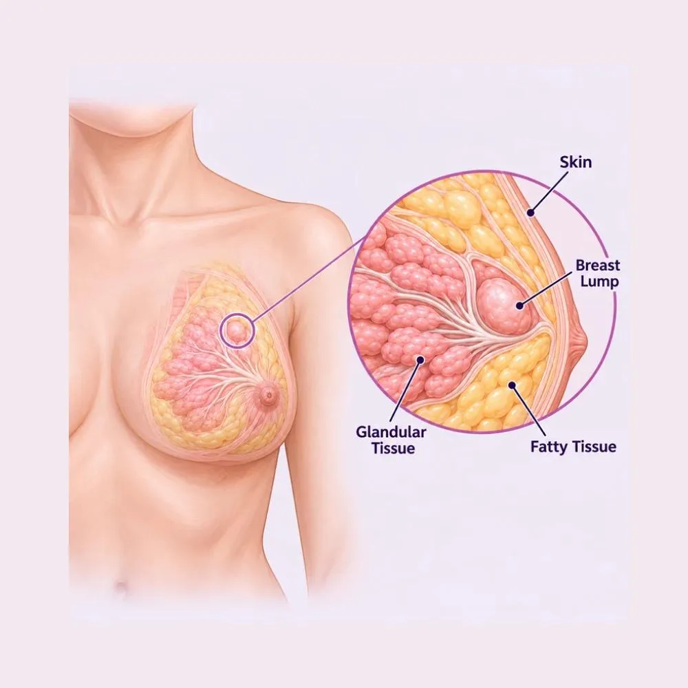
Breast lump surgery is a procedure to remove an abnormal lump from the breast tissue. It serves two purposes at once: it takes away the lump, and it provides tissue for examination under a microscope so the diagnosis is settled definitively.
Breast lumps fall into several categories. Benign lumps include fibroadenomas (smooth, mobile lumps common in younger women) simple cysts, lipomas and areas of fibrocystic change. Hormonal lumpiness fluctuates with the menstrual cycle. Infectious lumps arise from mastitis or an abscess. Malignant lumps are cancer, and are the reason every persistent lump gets properly assessed.
It is worth repeating that most lumps are benign. The purpose of assessment is not to find cancer in everyone but to identify the minority who have it, early enough that treatment is straightforward, while reassuring everyone else with certainty rather than with hope.
Most breast lumps are hormonal or structural changes in normal tissue. Cancer accounts for a small minority of lumps overall, though the proportion rises with age.
The commonest cause. Normal breast tissue responds to hormones with areas of lumpiness and tenderness, often worse before a period.
A benign, smooth, mobile lump made of glandular and fibrous tissue. Typical in women in their twenties and thirties, and entirely harmless.
Fluid-filled sacs that can appear suddenly and feel firm. Common around the forties, and easily confirmed and drained on ultrasound.
Menstruation, pregnancy, breastfeeding, menopause and hormonal contraception all change breast texture and can produce lumps.
Mastitis, most often during breastfeeding, can progress to an abscess: a painful, hot, tender lump needing drainage.
Injury to the breast can leave a firm scarred lump that mimics a tumour closely enough to need biopsy.
A soft, benign fatty lump, the same as those found elsewhere on the body.
Breast cancer, whose risk rises with age, family history, inherited gene mutations and hormonal factors. The minority that assessment exists to find.
The cause cannot be determined by feel alone, not by you, and not by an experienced surgeon either. That is precisely why imaging and a needle biopsy exist.
Knowing what is normal for your own breasts is what makes a change noticeable. These are the changes worth having checked.
Common findings
Changes needing prompt assessment
Any new lump that persists beyond one menstrual cycle should be assessed, whatever your age. So should any lump in a man: breast cancer in men is uncommon but not rare, and it is frequently diagnosed late.
The standard is triple assessment: clinical examination, imaging, and a needle biopsy. Together they are over ninety-nine per cent accurate, and most patients get their answer within days.
Assessment of the lump’s size, texture, mobility and fixation, along with the skin, nipple and the lymph nodes in the armpit.
The first-line imaging in women under forty, whose denser breast tissue makes mammography less useful. It distinguishes a cyst from a solid lump immediately.
X-ray imaging of the breast, used in women over forty. It detects microcalcification patterns that ultrasound cannot show.
A small tissue sample taken under local anaesthetic and ultrasound guidance. It gives a definitive tissue diagnosis, which no scan on its own can.
A fine needle samples cells, or drains a cyst, which often resolves the lump completely at the same appointment.
Reserved for selected cases: dense breasts, implants, an inherited high-risk gene, or where the other tests disagree with each other.
Where the three parts of triple assessment agree that a lump is benign, you can be genuinely reassured. Where they disagree, the lump is removed for certainty rather than watched.
Many lumps need no treatment beyond a confident diagnosis. Where treatment is needed, it ranges from draining a cyst in clinic to formal cancer surgery.
A small fibroadenoma or an area of fibrocystic change confirmed benign on triple assessment can simply be monitored, and many resolve on their own.
Draining a cyst with a fine needle in the clinic. The lump disappears immediately and no operation is needed.
For mastitis and breast abscesses, with ultrasound-guided aspiration often avoiding a formal surgical incision.
Adjusting hormonal contraception, and pain relief measures for cyclical breast pain and lumpiness.
A benign lump such as a fibroadenoma is removed through a single small probe under local anaesthetic, leaving a scar of a few millimetres.
The lump and a small margin of surrounding tissue are removed through a small incision. Both diagnostic and therapeutic, and the commonest breast operation.
For confirmed cancer, removing the tumour with a wider clear margin while preserving the rest of the breast. Usually followed by radiotherapy.
Removal of the whole breast, for larger or multifocal cancers, or where breast-conserving surgery is not appropriate.
The first lymph node draining the breast is identified and removed to check whether cancer has spread: sparing most patients a full armpit clearance.
Reshaping the remaining breast tissue, or reconstruction, so that the cosmetic result after cancer surgery is as good as it can be.
Surgery is advised for a persistent or growing lump, for severe pain or discomfort, where the diagnosis remains uncertain after triple assessment, and for any confirmed malignancy.
A thirty to sixty minute day-care procedure, usually under general anaesthetic, through an incision placed to hide in a natural crease or at the areolar edge.
Lumpectomy / excision biopsy
Triple assessment is completed first (examination, imaging and biopsy) so that the nature of the lump is known before, not after, the operation.
Where the lump is small or impalpable, a fine wire or marker is placed under ultrasound guidance beforehand so the surgeon can find it precisely.
General or local anaesthesia depending on size and site. The incision is planned along a natural skin line or at the areolar border, where scars fade best.
The lump is dissected out with a margin of surrounding tissue, taking care to preserve as much healthy breast as possible.
For cancer surgery, the specimen is X-rayed on the table to confirm the lesion and its marker are within it and the margins look adequate.
The breast tissue is reshaped to avoid leaving a dent, and the skin closed with a fine dissolvable subcuticular stitch.
Two to four hours in day care, then home the same day with painkillers, a supportive bra and a date to receive the histopathology result.
Every lump removed goes for histopathology without exception, even when the pre-operative biopsy said benign. The result is discussed with you in person at follow-up, usually within a week.
What matters most with a breast lump is a fast, definite answer. Here is why patients trust us:
Examination, imaging and needle biopsy together, so most patients get a definitive diagnosis rather than a series of appointments.
Consultants experienced in both benign breast surgery and oncoplastic cancer surgery, with attention to the cosmetic result.
From the first consultation through to post-treatment follow-up, with one team throughout.
Our hospital is in HITEC CITY, near IKEA: easily reachable from across Hyderabad.
Recovery after removal of a benign lump is quick: most patients are back to normal activities within a few days, with complete healing in a few weeks.
Home the same day. Some soreness and tightness at the site, controlled with oral painkillers. A supportive bra is worn day and night.
Most patients resume light normal activities. Bruising and swelling are expected and peak around day two.
Desk work resumes. Dressings come off and the wound is checked. Avoid raising the arm repeatedly overhead.
Bruising resolves and the wound settles. Histopathology is discussed at follow-up. Light exercise resumes; avoid high-impact activity.
Full activity including the gym. The scar softens and fades progressively, continuing to improve for up to a year.
Day and night for the first two weeks. Supporting the breast reduces swelling, discomfort and tension on the healing scar.
Take painkillers regularly rather than waiting for discomfort, and complete any antibiotic course prescribed.
No heavy lifting or strenuous activity for two to three weeks, and avoid repeated overhead arm movements early on.
Change dressings as instructed, shower rather than bathe for the first week, and pat the area dry.
Fever, spreading redness, increasing pain or swelling, discharge from the wound, or a rapidly enlarging firm swelling suggesting a haematoma.
This is where the histopathology result is discussed. Keep it even if you feel entirely well: the result is the point of the operation.
Keep checking your breasts afterwards. Removing one lump does not prevent another forming, and knowing your own breasts remains the most reliable way to notice a change early.
Surgery is advised where any of the following apply.
Cost depends on whether the lump is benign or malignant and on the procedure it requires. These are the ranges our patients fall into.
| Procedure | Estimated Cost |
|---|---|
| Consultation & clinical assessment Per visit | ₹800 – ₹1,500 |
| Ultrasound / mammography Out-patient | ₹1,500 – ₹5,000 |
| Core needle biopsy Including histopathology | ₹5,000 – ₹12,000 |
| Cyst aspiration Out-patient, done in clinic | ₹2,000 – ₹5,000 |
| Lumpectomy (benign lump) Day care, same-day discharge | ₹45,000 – ₹85,000 |
| Wide local excision with sentinel node Two to three nights’ stay | ₹1,50,000 – ₹2,80,000 |
| Mastectomy Three to five nights’ stay | ₹2,00,000 – ₹3,50,000 |
Removing a fibroadenoma is a day-care procedure. Cancer surgery involves node assessment, a longer stay and further treatment afterwards.
A deep or impalpable lump needs wire localisation beforehand, adding a step and a cost.
Triple assessment is standard. MRI and specialised receptor testing are added only where the case calls for them.
A shared ward, a twin-sharing room or a private suite: the largest single swing in most estimates.
We are empanelled with most major insurers, and our team files the pre-authorisation for you.
Every figure above is an indicative range, not a quote. After your consultation you are given a written, fixed-cost estimate, and that is the number you pay, with no additions once treatment begins.
What patients treated here can expect:
Triple assessment that settles the question within days, not weeks.
Incisions planned along natural lines, with minimally invasive options.
Diagnosis, surgery and follow-up from one team throughout.
The general and breast surgeons who assess and remove breast lumps at our HITEC CITY hospital.

Consultant General & Advanced Laparoscopic Surgeon
MBBS, MS, FMAS
17 years experience

Senior Consultant: Bariatric & Gastrointestinal Surgery
MBBS, MS, FRCS (UK), DLS (France)
28 years experience

Consultant Proctologist & Laparoscopic Surgeon
MBBS, MS, FMAS, FISCP, DMAS
8 years experience
Reviews from patients treated for breast, hernia and general surgery at Lux Hospitals, Hyderabad.
Written by Dr. Abhishek Katha on general and breast surgical care.

Dr. Abhishek Katha
Dr. Abhishek Katha

Dr. Abhishek Katha

Dr. Abhishek Katha

Dr. Abhishek Katha

Dr. Abhishek Katha
Common questions about breast lumps, their removal, and what treatment costs at Lux Hospitals.
Still have questions? Call us at 079 6908 4422
Still have questions? Message us on WhatsApp at 079 6908 4444
Triple assessment for a fast, definite answer, with a fixed-cost estimate before any treatment begins.
Plot No.116, Lumbini Avenue, HITEC CITY, Near IKEA, Hyderabad – 500032