
Dr. Samhitha Reddy
Consultant Proctologist & Laparoscopic Surgeon
MBBS, MS, FMAS, FISCP, DMAS
- 8 Yrs
- ASCRS Member
Fibroids are benign muscular growths in the wall of the uterus, and they are extremely common: by the age of fifty, most women have had one. Many never know.
The ones that matter cause heavy bleeding, pressure and pain, and they are frequently treated with a hysterectomy that was not necessary. Our gynaecologists at Lux Hospitals, HITEC CITY begin from the position that the uterus is worth preserving where it reasonably can be.
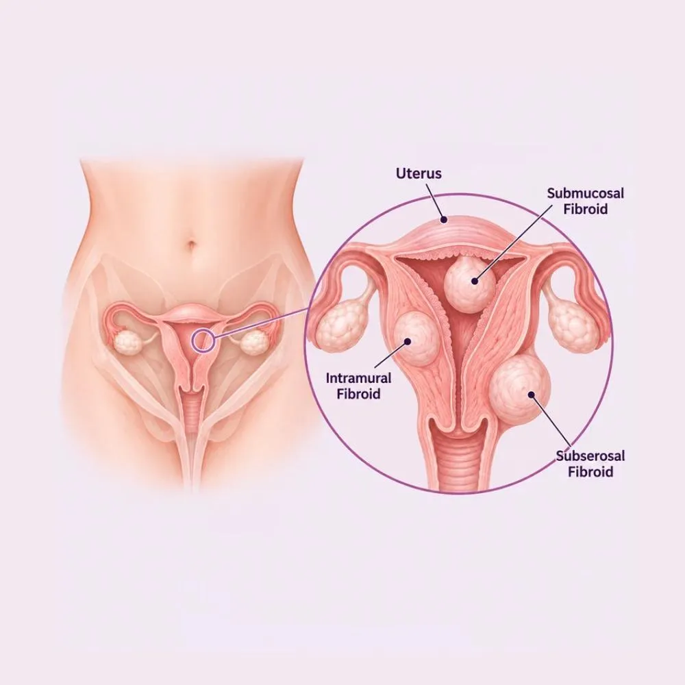
Fibroids (properly leiomyomas) are benign tumours arising from the smooth muscle of the uterine wall. Each grows from a single cell, which is why a woman can have one fibroid or twenty, ranging from a few millimetres to the size of a melon. They are not cancer, and they do not become cancer: the malignant equivalent, leiomyosarcoma, is a distinct and rare tumour that arises independently.
Where a fibroid sits matters far more than how big it is. Submucosal fibroids bulge into the cavity of the uterus and cause heavy bleeding and miscarriage out of all proportion to their size. Intramural fibroids sit within the muscle wall and are the commonest type. Subserosal fibroids project outward and tend to cause pressure on the bladder or bowel rather than bleeding. Pedunculated fibroids hang on a stalk and can twist.
Fibroids are oestrogen and progesterone dependent. They grow through the reproductive years, often enlarge during pregnancy, and characteristically shrink after menopause. This hormonal dependence is what makes several of the medical treatments work, and it is also why a fibroid growing after menopause is treated as a warning sign rather than a nuisance.
The trigger for the initial cell change is not known, but the factors that drive growth are well established.
Fibroids carry more hormone receptors than surrounding muscle. They grow through the reproductive years and shrink after menopause.
Specific genetic changes are found within fibroid tissue, and a mother or sister with fibroids substantially raises your risk.
Fibroids are considerably commoner, appear earlier and grow larger in women of African descent: one of the strongest associations known.
Incidence rises through the thirties and forties and peaks in the years before menopause.
Fat tissue produces oestrogen, and higher body weight is associated with both more fibroids and larger ones.
Pregnancy appears protective, and risk falls with each pregnancy carried to term.
Consistently associated with fibroid development, and worth correcting though it is not established as a treatment.
Nothing in your diet or lifestyle caused your fibroids. Diet influences growth modestly at most, and claims that particular foods dissolve fibroids have no basis.
Most fibroids cause nothing. Where they do, the symptoms follow from where they sit.
Bleeding symptoms
Pressure and other symptoms
Heavy bleeding is the symptom most often endured for years. Iron-deficiency anaemia from fibroids is extremely common and easily missed, and correcting it frequently transforms how a woman feels regardless of what is done about the fibroid.
Ultrasound diagnoses most fibroids. Further imaging is about mapping them precisely enough to plan treatment.
An enlarged, firm, irregular uterus is often felt. Frequently the first indication, and sometimes found incidentally.
The first-line test. It counts the fibroids, measures them and (most importantly) establishes their position relative to the cavity.
Fluid instilled into the cavity to outline submucosal fibroids, which are the ones causing heavy bleeding and fertility problems.
Maps large or numerous fibroids precisely before myomectomy or embolisation, and helps distinguish fibroids from adenomyosis.
Haemoglobin and ferritin, since chronic heavy bleeding causes iron deficiency long before it causes obvious anaemia.
Direct inspection of the cavity, which both confirms submucosal fibroids and allows them to be removed in the same procedure.
A fibroid that grows after menopause, or that grows unusually rapidly, is investigated further. It is very rarely sinister, but that is precisely the situation where it must not be assumed benign.
Treatment depends on your symptoms, your fibroids’ position and whether you want to preserve fertility, not on their size alone.
Correct for fibroids causing no symptoms, whatever their size. They frequently shrink after menopause without any intervention.
Reduce menstrual blood loss substantially without affecting the fibroid itself. Taken only during the period.
A levonorgestrel device that markedly reduces bleeding. Well suited to intramural fibroids where the cavity is not distorted.
Shrink fibroids by inducing a temporary menopausal state. Used before surgery to reduce size and correct anaemia, not long term.
Oral or intravenous iron to correct the anaemia, which often improves symptoms as much as treating the fibroid does.
A radiologist blocks the arteries supplying the fibroids through a catheter in the wrist or groin. No incision, short recovery, uterus preserved.
Submucosal fibroids removed through the cervix with no incision at all. Day care, and the treatment of choice for bleeding and fertility problems.
Keyhole removal of intramural and subserosal fibroids with the uterus repaired and preserved. Small incisions and a faster recovery than open surgery.
Through an abdominal incision, for very large or numerous fibroids where keyhole removal is not feasible.
Removal of the uterus where family is complete and other treatments have failed. Definitive, and the only option that guarantees no recurrence.
Destroying the uterine lining to control bleeding, suitable for small fibroids where the cavity is not distorted and family is complete.
Hysterectomy is the only treatment that eliminates the possibility of recurrence, but for a woman who wants to keep her uterus, myomectomy and embolisation both work well, and the choice belongs to her.
Myomectomy takes ninety minutes to three hours depending on the number and position of the fibroids.
Keyhole uterus-preserving surgery under general anaesthesia
Anaemia corrected with iron beforehand, and GnRH analogues sometimes given for two to three months to shrink large fibroids and reduce blood loss.
General anaesthesia, with a camera port at the umbilicus and three further small ports across the lower abdomen.
Vasopressin injected into the uterine wall around each fibroid to constrict vessels. Blood loss is the main technical challenge of this operation.
An incision made over the fibroid and carried down to its capsule: the plane where it separates cleanly from normal muscle.
The fibroid shelled out of its capsule with traction and countertraction, staying in the correct plane so healthy muscle is preserved.
The defect closed in layers with strong sutures. This repair is what determines whether the uterus is safe for a future pregnancy.
Fibroids removed in a containment bag, an anti-adhesion barrier placed, and the ports closed.
The layered closure of the uterine wall is the step that matters most for anyone hoping to conceive. A poorly repaired uterus carries a genuine risk of rupture in labour, and it is the reason this operation rewards experience.
Too many fibroids are treated with hysterectomy when the uterus could have been kept. Here is why patients trust us:
Hysteroscopic and laparoscopic myomectomy offered as genuine first options, with hysterectomy discussed rather than assumed.
Where a fibroid sits determines what it causes and how it is best removed, so it is mapped properly rather than treated on size alone.
From the first consultation through to post-treatment follow-up, with one team throughout.
Our hospital is in HITEC CITY, near IKEA: easily reachable from across Hyderabad.
Recovery ranges from a same-day discharge after hysteroscopic removal to three weeks after open myomectomy.
Hysteroscopic myomectomy is day care with light bleeding for a few days. After laparoscopic myomectomy, discharge at two to three days.
Port-site soreness settles. Light bleeding or discharge is expected. Most women return to desk work at two weeks.
Activity increases gradually. Intercourse resumes at four to six weeks. Histology of the removed fibroids is reviewed at follow-up.
Bleeding patterns settle and anaemia corrects. After myomectomy, conception is generally advised to wait three to six months.
Periodic ultrasound where fibroids remain or recur. Recurrence after myomectomy is common: roughly a quarter within five years.
Three to six months allows the uterine repair to heal fully. Conceiving too soon risks the scar giving way later in pregnancy.
Years of heavy bleeding deplete iron stores well beyond what haemoglobin shows. Replacing it often improves how you feel more than anything else.
A myomectomy scar in the uterine wall affects how labour is managed, and a caesarean is often recommended. This must be on record.
Myomectomy removes existing fibroids but not the tendency to form them. Around a quarter of women develop new ones within five years.
The uterine repair and the abdominal wall both need time. Walking is encouraged from the first day.
Bleeding heavier than a normal period, fever, or increasing rather than settling pain all need review rather than waiting.
Fibroids shrink naturally after menopause. For a woman close to it with tolerable symptoms, waiting is a legitimate strategy rather than avoidance, and it is worth discussing openly.
Book a consultation if any of the following apply.
Cost varies widely with the treatment chosen, and several effective options avoid surgery entirely.
| Procedure | Estimated Cost |
|---|---|
| Consultation Per visit | ₹800 – ₹1,500 |
| Transvaginal ultrasound Counts and maps fibroids | ₹1,500 – ₹3,500 |
| MRI pelvis Before myomectomy or embolisation | ₹8,000 – ₹18,000 |
| Hormonal IUD insertion Includes device; lasts five years | ₹12,000 – ₹25,000 |
| Hysteroscopic myomectomy Day care; no incision | ₹75,000 – ₹1,40,000 |
| Laparoscopic myomectomy Two to three nights | ₹1,50,000 – ₹2,80,000 |
| Uterine fibroid embolisation Includes embolic material | ₹1,80,000 – ₹3,20,000 |
| Laparoscopic hysterectomy Two to three nights | ₹1,60,000 – ₹2,90,000 |
A single submucosal fibroid is removed hysteroscopically as day care. Multiple large intramural fibroids require a considerably longer operation.
Myomectomy is technically more demanding than hysterectomy and takes longer, but it keeps the uterus, which for many women is the point.
Correcting anaemia and shrinking fibroids with GnRH analogues beforehand adds cost but makes surgery safer.
A shared ward, a twin-sharing room or a private suite: the largest single swing in most estimates.
We are empanelled with most major insurers, and our team files the pre-authorisation for you.
Every figure above is an indicative range, not a quote. After your consultation you are given a written, fixed-cost estimate, and that is the number you pay, with no additions once treatment begins.
The gynaecologists who treat uterine fibroids at our HITEC CITY hospital.

Consultant Gynaecologist & Advanced Laparoscopic Surgeon
MBBS, MS, FMAS
16 years experience

Consultant Obstetrician, Gynecologist & Laparoscopic Surgeon
MBBS, MS (Obstetrics & Gynaecology)
11 years experience
Reviews from women treated for gynaecological conditions at Lux Hospitals, Hyderabad.
Written by Dr. Harshita Kakarla on women’s health and gynaecological care.
Dr. Harshita Kakarla
Dr. Harshita Kakarla
Dr. Harshita Kakarla

Dr. Harshita Kakarla
Dr. Harshita Kakarla
Dr. Harshita Kakarla
Common questions about uterine fibroids, myomectomy, hysterectomy and costs at Lux Hospitals.
Still have questions? Call us at 079 6908 4422
Still have questions? Message us on WhatsApp at 079 6908 4444
Fibroids mapped properly and treated with the uterus preserved wherever possible, with costs shared upfront.
Plot No.116, Lumbini Avenue, HITEC CITY, Near IKEA, Hyderabad – 500032