
Dr. Samhitha Reddy
Consultant Proctologist & Laparoscopic Surgeon
MBBS, MS, FMAS, FISCP, DMAS
- 8 Yrs
- ASCRS Member
The shoulder is the most mobile joint in the body, and that mobility comes at the cost of stability: it dislocates more often than any other joint. A first dislocation is intensely painful and needs putting back promptly.
What happens afterwards matters more than most people realise. Our orthopaedic surgeons at Lux Hospitals, HITEC CITY reduce dislocations urgently, image the damage properly, and offer arthroscopic stabilisation where the risk of it happening again is high.
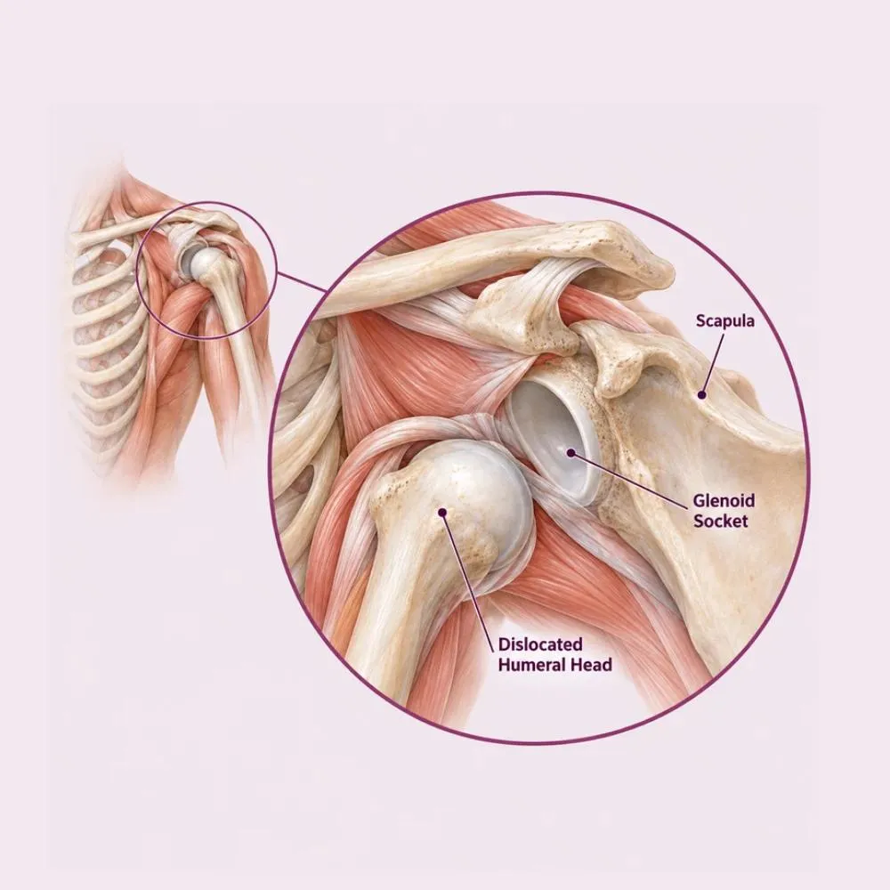
Shoulder dislocation occurs when the head of the humerus comes completely out of the shallow socket of the shoulder blade. The socket is genuinely shallow, roughly the depth of a golf tee holding a golf ball, and stability depends on the surrounding labrum, capsule and muscles rather than on bone.
Over ninety per cent of dislocations are anterior, with the ball coming out forward, typically from a fall on an outstretched arm or a forced external rotation. Posterior dislocations are much less common and are classically associated with seizures and electric shocks, and are frequently missed on a single X-ray view. Inferior dislocations are rare.
The important consequence is what tears in the process. The labrum is usually pulled off the front of the socket (a Bankart lesion) and once that has happened the shoulder is structurally more likely to dislocate again. This is why the risk of recurrence after a first dislocation is so high in young people.
Most dislocations follow a specific injury, but who dislocates again afterwards depends heavily on age and activity.
The commonest mechanism. Force transmitted up the arm levers the humeral head forward out of the socket.
Kabaddi, rugby, hockey and football all involve tackles and falls with the arm in the vulnerable position.
The arm being pulled or twisted outward with the elbow away from the body: the classic anterior dislocation position.
High-energy trauma that may dislocate the shoulder alongside fracturing it, which changes management considerably.
Violent muscle contraction drives the shoulder backward, causing posterior dislocation: the type most often missed.
The single strongest predictor of recurrence. Under twenty, the risk of dislocating again exceeds seventy per cent.
Naturally loose joints, sometimes as part of a hypermobility syndrome, predispose to instability with minimal trauma.
Each dislocation can chip bone from the socket rim, and beyond a certain amount the shoulder cannot be stabilised by soft tissue repair alone.
Age is the decisive factor. A twenty-year-old athlete who dislocates has a very high chance of it happening again; a sixty-year-old often does not, but is far more likely to have torn the rotator cuff instead.
An acute dislocation is unmistakable. The pattern afterwards (recurrent instability) is what brings people back.
At the time of dislocation
Ongoing instability afterwards
Numbness down the arm or a cold, pale hand after a dislocation needs urgent attention. The axillary nerve is commonly stretched, and vascular injury, though rare, is an emergency.
The dislocation itself is diagnosed clinically and confirmed on X-ray. What matters afterwards is imaging the damage that was done.
The characteristic deformity and loss of movement, with careful assessment of the axillary nerve and the pulse in the arm.
Confirm the direction of dislocation and exclude fracture, then confirm the joint is correctly relocated. Two views are essential to avoid missing a posterior dislocation.
Shows the labral tear, capsular damage and any associated rotator cuff tear. An arthrogram with contrast is most sensitive for labral injury.
Quantifies bone loss from the socket rim and any impaction fracture on the humeral head, which determines whether soft tissue repair will suffice.
Apprehension and relocation tests performed once the acute episode has settled, assessing the direction and degree of instability.
Anyone over forty who dislocates is checked for a rotator cuff tear, which is common in that group and easily overlooked.
Bone loss from the socket rim is the finding that changes the operation. Beyond around twenty per cent, a soft tissue repair will fail and a bone block procedure is needed instead.
The immediate treatment is reduction. What follows depends on your age, your activities and what the imaging shows.
Putting the joint back, under sedation and muscle relaxation. Doing it promptly reduces pain immediately and limits further damage.
A sling for two to three weeks after a first dislocation, allowing the torn capsule and labrum to begin healing.
Strengthening the rotator cuff and the muscles controlling the shoulder blade, which improves dynamic stability considerably.
Avoiding the provocative position, arm out and rotated outward, during the rehabilitation phase.
Analgesia and ice in the acute phase, which is when discomfort is at its worst.
The torn labrum is reattached to the socket rim with suture anchors through keyhole portals. The standard operation for recurrent instability.
Tightening a stretched capsule alongside the labral repair, particularly where there is generalised laxity.
Transferring a piece of the coracoid bone with its attached tendon to the front of the socket. The operation for significant bone loss, where soft tissue repair would fail.
Filling a large impaction defect on the humeral head with cuff tissue, preventing it engaging on the socket rim.
Grafting bone to rebuild a deficient socket rim, for revision cases or severe bone loss.
In patients over forty, repairing the cuff tear that frequently accompanies dislocation in that age group.
Early stabilisation after a first dislocation is increasingly offered to young athletes, because the recurrence risk in that group is so high and each further dislocation causes more bone loss.
A sixty to ninety minute arthroscopic procedure through three or four small portals, usually as a day case.
Arthroscopic Bankart repair
MRI and CT reviewed to confirm the labral tear and quantify bone loss, which determines whether a Bankart repair or Latarjet is appropriate.
General anaesthesia with an interscalene nerve block for post-operative pain relief.
The joint is inspected, the labral tear confirmed, and the rotator cuff and cartilage assessed at the same time.
The torn labrum is freed and the bony rim lightly abraded to a bleeding surface, so the labrum can heal back to it.
Suture anchors are inserted into the socket rim (typically three or four) each loaded with strong sutures.
The labrum is drawn back onto the rim and secured, restoring the bumper that deepens the socket, and the capsule tightened as needed.
Portals closed, arm placed in a sling, and home the same day with a staged rehabilitation plan.
Where CT shows significant bone loss, a Latarjet is done instead: transferring bone to deepen the socket. Choosing a soft tissue repair in the face of bone loss is the commonest reason stabilisation fails.
What happens after the first dislocation determines whether there is a second. Here is why patients trust us:
Prompt reduction under sedation with proper imaging before and after, which limits further damage and relieves pain immediately.
MRI and CT to quantify labral injury and bone loss, so the right stabilisation procedure is chosen rather than the default one.
From the first consultation through to post-treatment follow-up, with one team throughout.
Our hospital is in HITEC CITY, near IKEA: easily reachable from across Hyderabad.
After a first dislocation treated without surgery, recovery takes six to twelve weeks. After stabilisation surgery it is four to six months.
After reduction, the pain settles dramatically. Sling for comfort, with ice and analgesia.
Sling worn to allow the capsule to heal. Gentle pendulum exercises only, avoiding the provocative position.
Sling discarded. Progressive range of motion, avoiding external rotation with the arm abducted.
Strengthening the rotator cuff and scapular muscles. Return to non-contact activity and desk work.
Return to contact sport after surgical stabilisation, guided by strength, control and confidence rather than time alone.
Arm out to the side and rotated outward is the position of instability. Avoid it during rehabilitation, and be careful with it long term.
Dynamic stability comes from these muscles. They are what compensate for a socket that is shallow by design.
After surgery it protects the repair while the labrum heals to bone. After a first dislocation it allows the capsule to tighten.
Numbness or weakness in the arm, a cold or pale hand, fever or wound discharge after surgery, or the shoulder coming out again.
Returning before strength and control are restored is the main cause of re-dislocation, and each episode causes more bone loss.
Ongoing cuff and scapular strengthening reduces the risk of further instability, particularly in contact athletes.
Each further dislocation chips more bone from the socket rim. Beyond a certain point a simple soft tissue repair will no longer hold, and a bigger bone block operation becomes necessary, which is the argument for stabilising early in high-risk patients.
A dislocated shoulder is an emergency: come in immediately for the first three.
Emergency reduction is inexpensive. Stabilisation surgery is the significant cost, and only some patients need it.
| Procedure | Estimated Cost |
|---|---|
| Consultation & assessment Per visit | ₹800 – ₹1,500 |
| Emergency reduction under sedation Including X-rays before and after | ₹12,000 – ₹30,000 |
| MRI or MR arthrogram Assesses labral and cuff damage | ₹8,000 – ₹18,000 |
| CT scan for bone loss Where recurrent dislocation has occurred | ₹5,000 – ₹12,000 |
| Arthroscopic Bankart repair Day care or one night; anchors included | ₹1,30,000 – ₹2,40,000 |
| Latarjet procedure One to two nights’ stay | ₹1,80,000 – ₹3,20,000 |
| Post-operative physiotherapy Four to six months of rehabilitation | ₹15,000 – ₹35,000 |
A Bankart repair uses anchors; a Latarjet involves bone transfer with screws and is a longer, more complex operation.
Typically three or four for a standard repair, more where the labral tear is extensive.
A rotator cuff tear in an older patient, or a fracture, adds to the procedure and the cost.
A shared ward, a twin-sharing room or a private suite: the largest single swing in most estimates.
We are empanelled with most major insurers, and our team files the pre-authorisation for you.
Every figure above is an indicative range, not a quote. After your consultation you are given a written, fixed-cost estimate, and that is the number you pay, with no additions once treatment begins.
The orthopaedic and shoulder surgeons who treat shoulder instability at our HITEC CITY hospital.

Consultant Orthopaedic Surgeon: Joint Replacement & Arthroscopy
MS (Ortho), FIJR, FIASM (UK), FIKS (Italy), FIRJR
8 years experience

Consultant Orthopaedic Surgeon
MBBS, MS, FIJR
6 years experience
Reviews from patients treated for shoulder, knee and sports injuries at Lux Hospitals, Hyderabad.
Written by Dr. Sai Kishan Sirasala on shoulder injuries, instability and orthopaedic care.
Dr. Sai Kishan Sirasala
Dr. Sai Kishan Sirasala
Dr. Sai Kishan Sirasala
Dr. Sai Kishan Sirasala
Dr. Sai Kishan Sirasala
Dr. Sai Kishan Sirasala
Common questions about shoulder dislocation, stabilisation surgery, and what it costs at Lux Hospitals.
Still have questions? Call us at 079 6908 4422
Still have questions? Message us on WhatsApp at 079 6908 4444
Urgent reduction and full assessment of the damage, with stabilisation surgery where the risk of recurrence is high.
Plot No.116, Lumbini Avenue, HITEC CITY, Near IKEA, Hyderabad – 500032